 Jennifer Mathis, DVM, DAVDC, CVPP
1 min read
Treating Periodontal Disease: Ziggy's Journey with Guided Tissue Regeneration
Case Summary: Guided Tissue Regeneration (GTR) is a technique used to promote the regeneration of bone and soft tissue, enabling the replacement of...
Guided Tissue Regeneration (GTR) is a technique performed to promote osseous regeneration, allowing for tissue growth (bone and soft tissues) to replace an infrabony periodontal pocket (a site of vertical bone loss). This involves cleaning the infrabony pocket using hand curettes, potentially with a specialized ultrasonic curette as well. A bone graft, along with a barrier membrane, is placed into the pocket, followed by tight closure of the gingival collar. GTR is performed to preserve structural integrity in cases of vertical bone loss.
Buffy, a 6yo FS 11# Beagle presented from the referring veterinarian (RDVM), Dr. Ryan Southard, at Family Pet Veterinary Center, for a Comprehensive Oral Health Assessment and Treatment (COHAT). Initial exam findings calculus index 1-3 in varied areas of the mouth; 409 gingival recession (GR) 4mm with exudate at the furcation.
PD2 (Periodontal Disease Index <25% attachment loss) 102-103, 202-203, 104
PD3 (Periodontal Disease Index 25-50% attachment loss) 204
FE2 (Furcation Exposure under the crown) GR (Gingival Recession) 409, 4mm
Missing 105-109, 201, 205-207, 306-308, 302-303, 401-403, 405, 408
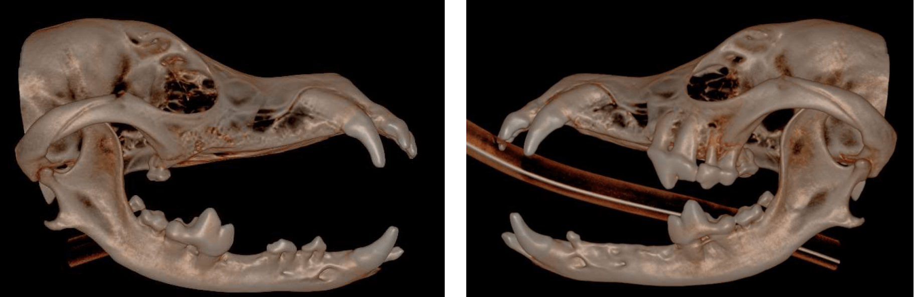
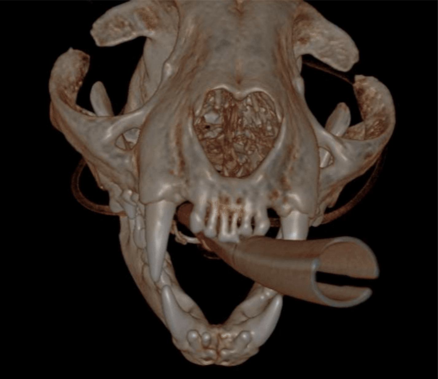
Below: CBCT Hard tissue reconstruction
Below: High intensity CBCT Image
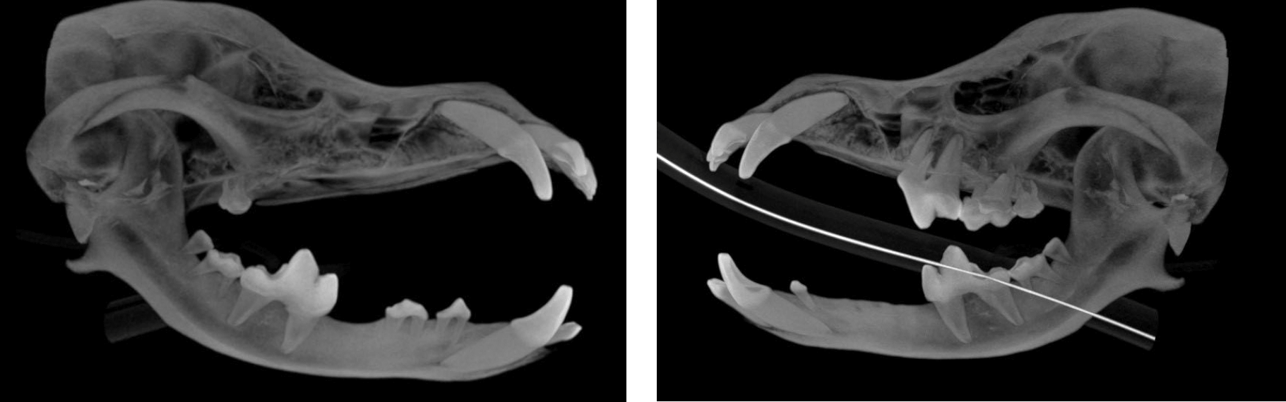
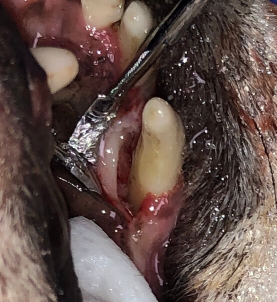
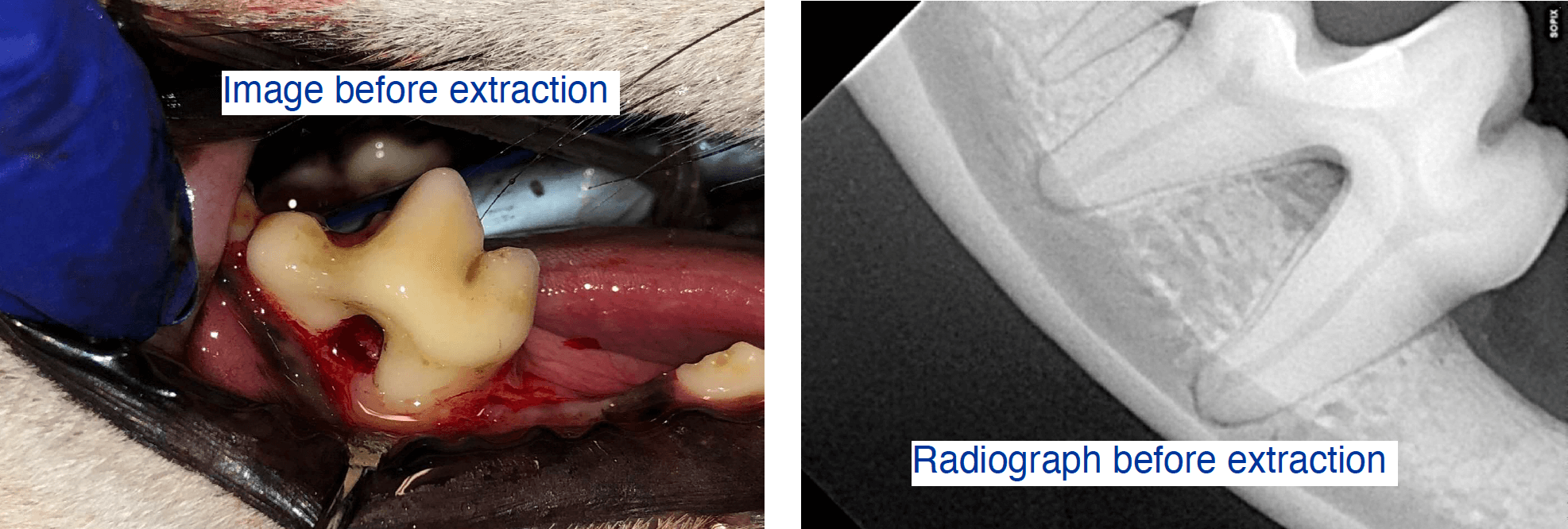
Below: Bone loss affecting upper left canine tooth
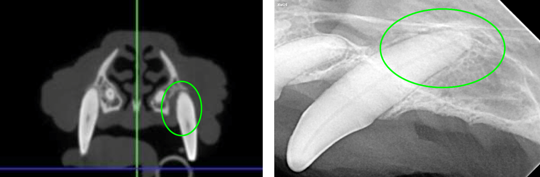
Below: Imaging of both sides of the lower right 4th premolar
Furcation exposure level 2 indicating bone loss under the crown
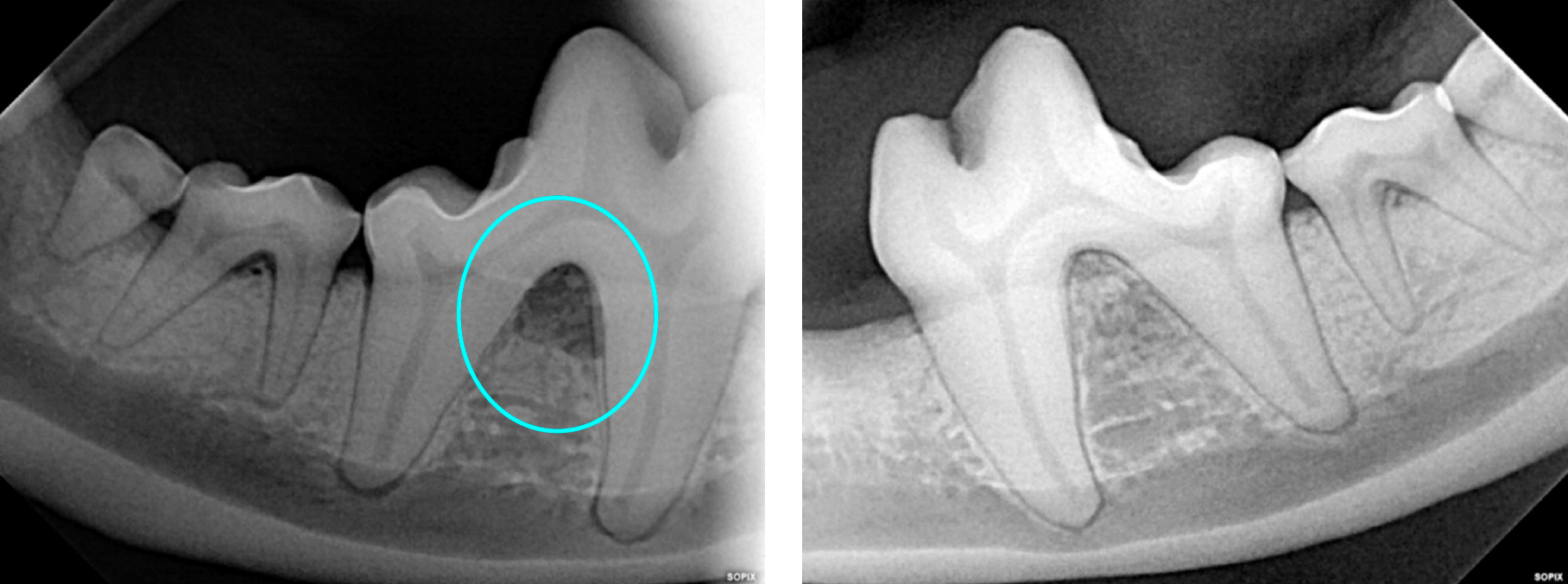
During the dental procedure Dr. Ryan noted probing depth of 8mm at the palatal aspect 204 and 5mm palatal probing depth of 104. Closed root planing (RP/C) was performed at this time and referred for Guided Tissue Regeneration (GTR).
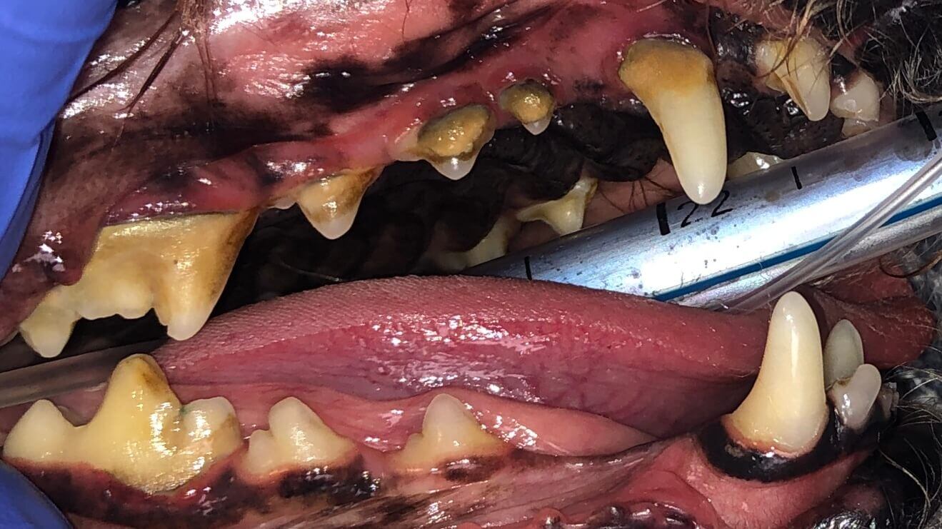
Below: Presentation at the beginning of procedure with RDVM
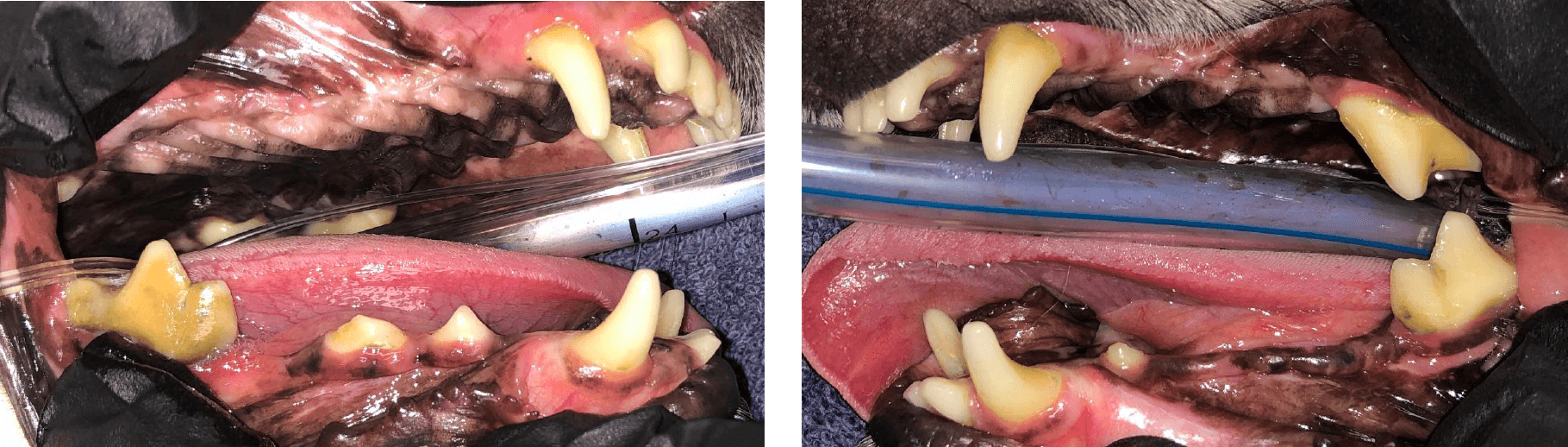
Below: After procedure with RDVM (Before referral to Animal Dentistry Referral Services)
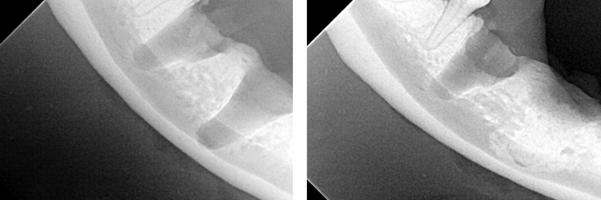
In the one month after the referring DVM performed closed root planing, the upper right canine palatal pocket had improved from 5mm to 3mm (within normal limits) and the upper left canine infra-bony pocket depth had also improved from 8mm to 6mm. It’s typical to have a 1-2mm improvement in probing depth in the short term (1-3 months). Closed root planing turns an active periodontal pocket inactive. Recurrence of active periodontal disease is dependent up on pocket depth and dental home care as well as sooner (3-6 months) professional anesthetic dental procedures.
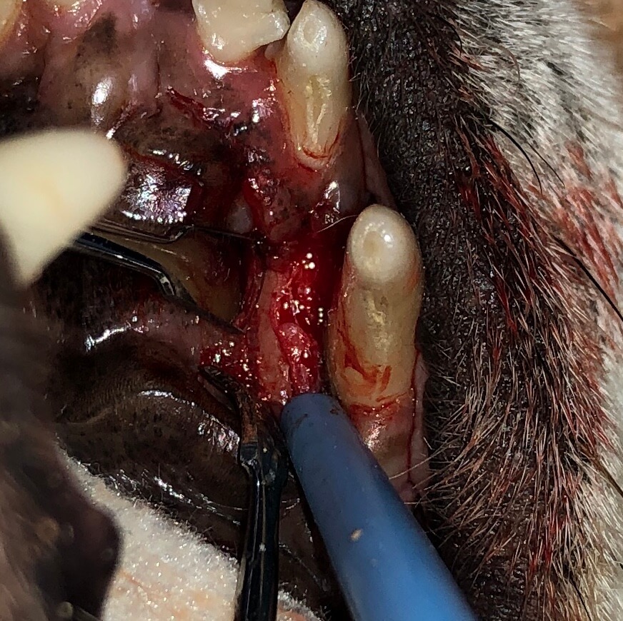
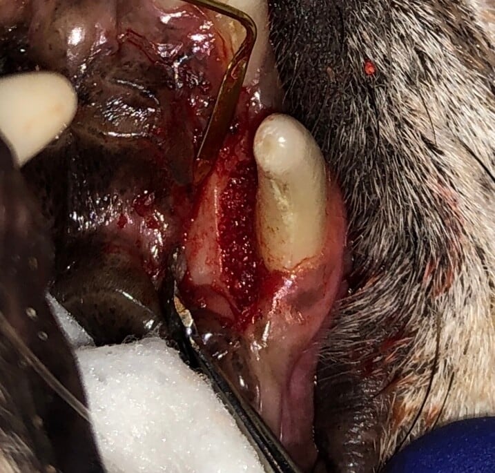
An envelope flap was utilized, followed by curettage to remove granulation tissue and debris. Osteoallograft periomix bone graft material from Veterinary Transplant Services was then placed in the infrabony pocket. Doxirobe was spread thinly to create a membrane covering the graft material, and closure was achieved using 4-0 monocryl to create a tight gingival collar.
The images below were taken at various stages throughout the procedure.
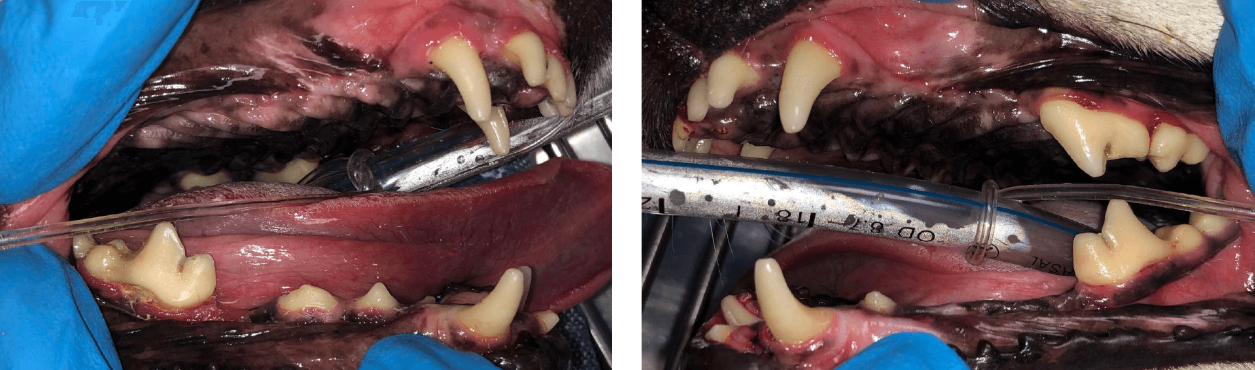
Showing deep bone loss at palatal aspect of upper left canine tooth before an oral nasal fistula has developed

View of the granulation bed which must be fully removed to be able to treat
Also showing some debris, a good lesson to use magnification and clean more
Mixed with blood during GTR procedure
To prevent soft tissue down growth and the bone returns
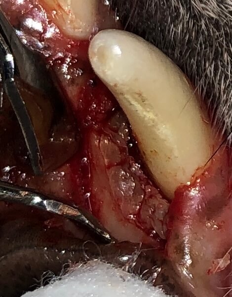
With tight gingival collar

Envelope flap, section, elevate, graft placement. The site was closed with4-0 monocryl.
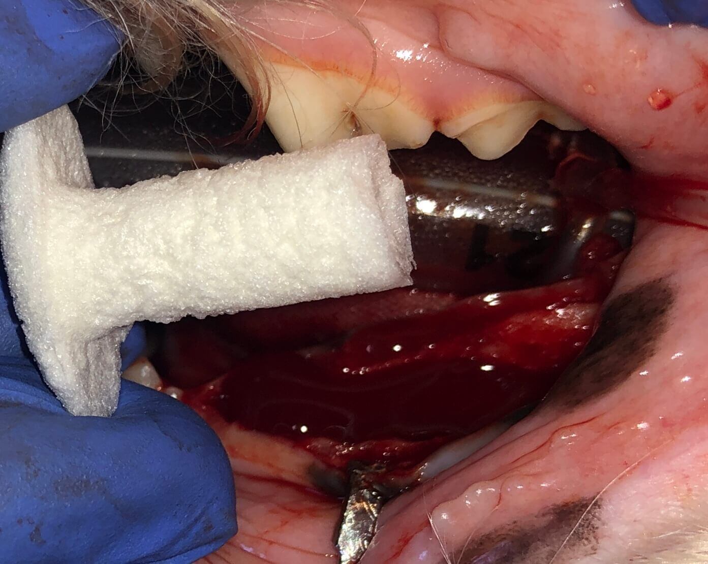
Below: (left) BEFORE extraction, (right) AFTER extraction
Buffy is a regular client and is doing great. Owner has no concerns and continues to see us regularly for the bone graft study. The most recent dental examination under anesthesia was September of 2023. Dental procedure findings: PP 0-1m 104, 204 with no bleeding on probing ie GTR is looking great, ie bone has returned to the area and attachment loss has been regained. Next dental procedure scheduled June 2024.
1 min read
Case Summary: Guided Tissue Regeneration (GTR) is a technique used to promote the regeneration of bone and soft tissue, enabling the replacement of...
1 min read
Case Summary: Stomatitis refers to widespread inflammation of the mucous lining of the mouth's structures, often associated with an underlying immune...
1 min read
Case Summary & Patient Information: Daisy, a 2.5-year-old spayed female Old English Sheepdog/Poodle mix, presented with a primary complaint of a lump...