1 min read
Complicated Dental Fractures: The Benefits of Root Canal Therapy
Case Summary: In this case study, we delve into the intricate treatment of a complicated crown fracture in a canine tooth, where the pulp is exposed,...
2 min read
 Jennifer Mathis, DVM, DAVDC, CVPP
:
Oct 23, 2024 1:16:12 PM
Daisy, a 2.5-year-old spayed female Old English Sheepdog/Poodle mix, presented with a primary complaint of a lump under her left mandible. This lump was first noted by the referring veterinarian (RDVM) on January 30, 2024. According to the owner, the lump had been present for a couple of weeks and had changed in size. During the exam with the RDVM, Daisy showed discomfort when the lump was palpated.
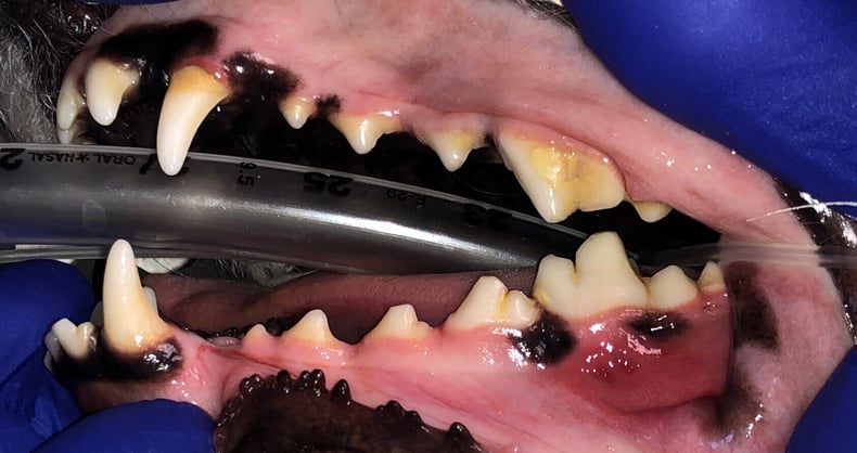
The owner also mentioned that Daisy had a history of kennel chewing and had experienced trauma from kenneling in 2022-2023, including an avulsed premolar. Due to the complexity of the case, Daisy was referred to Dr. Jennifer Mathis, DAVDC, at Animal Dentistry Referral Services for a cone-beam computed tomography (CBCT) scan and further treatment.
A firm swelling was detected on the left mandibular region. While the exact cause is unclear, it is suspected that trauma occurring between 10 and 24 months of age led to the death of the lower molar. Another possible cause is Mandibular Periostitis Ossificans (MPO), a developmental condition seen in large breed dogs. In MPO cases, the swelling typically resolves on its own. However, Daisy’s condition, if MPO, would extend beyond the usual timeline, making trauma the more likely cause.
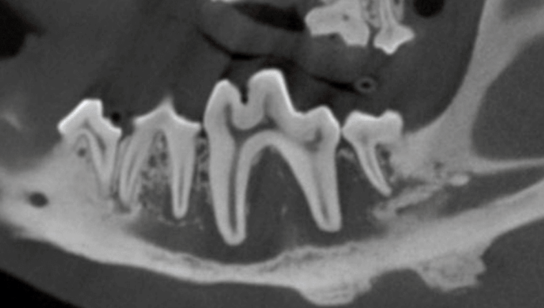
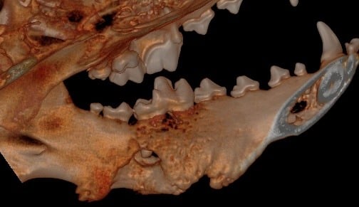
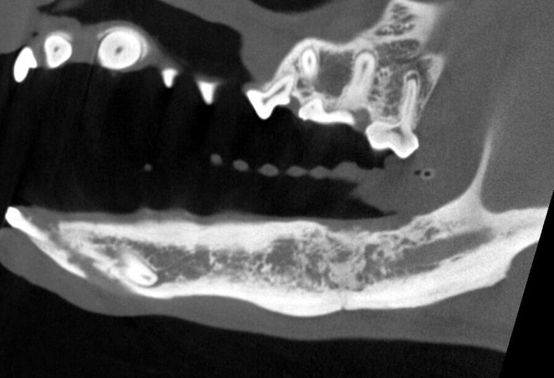
The internal structure of the affected tooth showed disease at the root tips, leading to infection. This infection had spread into the mandibular canal and affected nearby teeth. Although there was reactive bony proliferation on the ventral aspect, significant bone loss was also noted, which posed a risk for a pathological mandibular fracture.
Below: CT Cross Section of Disease
Below: 3D Hard Tissue Overview (left side)
.jpg?width=790&height=486&name=3D%20Hard%20Tissue%20Overview%20(left%20side).jpg)
.jpg?width=629&height=390&name=3D%20Hard%20Tissue%20Overview%20(left%20mandible).jpg)
Below: 3D Hard Tissue Lingual View (left mandible)
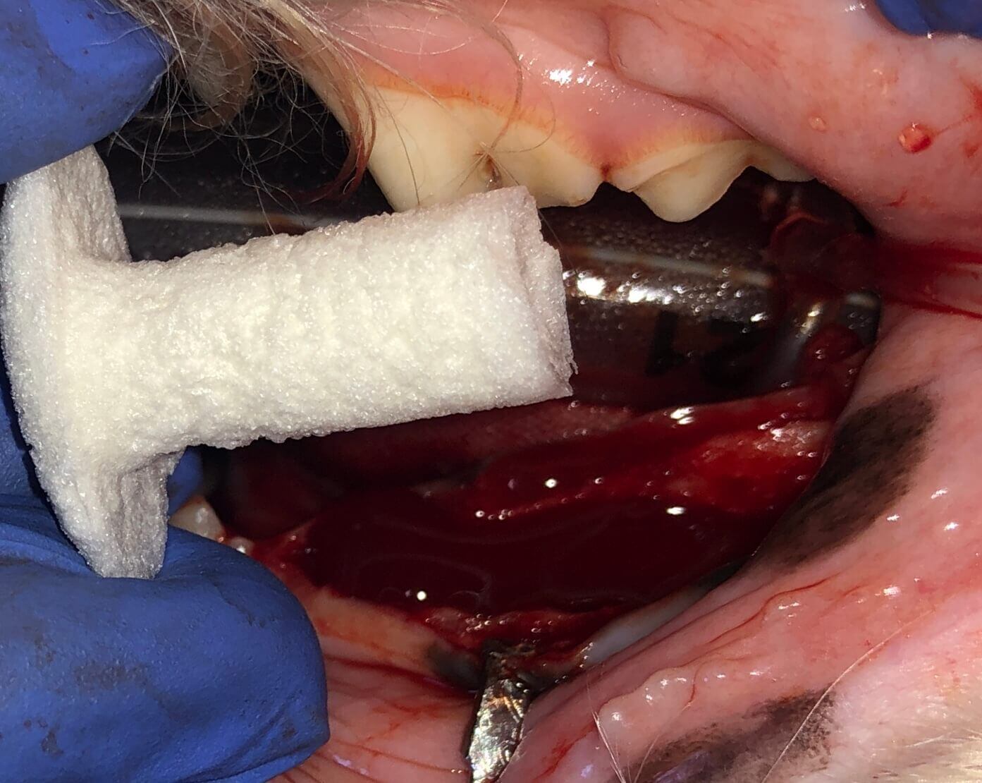
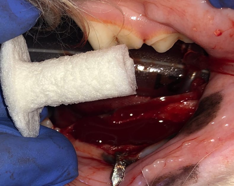
The diseased teeth were surgically extracted, and the site was cleaned down to healthy bone. A bone scaffold and membrane (ReGum Vet "mushroom" or "plug") were placed to aid in mandibular healing. ReGum is composed of a cross-linked gelatin foam matrix. It fills the defect and supports the tissue. One of the benefits of ReGum Vet is that it requires no suturing.
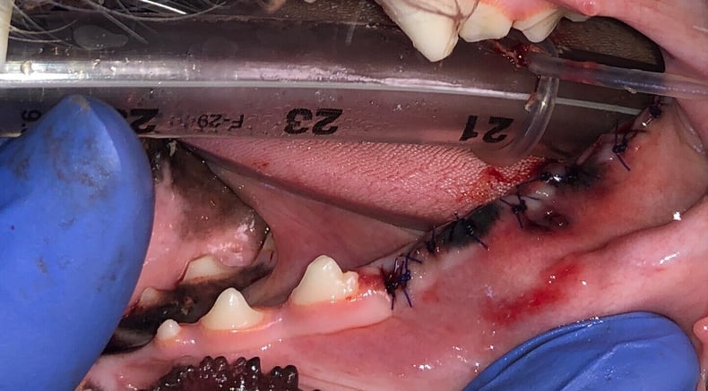
After thorough cleaning, the ReGum product is placed and allowed to absorb blood, functioning like a sponge. Once it has soaked adequately, the surgical site is sutured closed. Pro tip: Prepare the surgical site for closure before placing ReGum to avoid dislodging it during flap trimming.
In Daisy's case, a culture sample was taken prior to cleaning. The results identified Pasteurella species, Moraxella species, and Staphylococcus pseudintermedius. Daisy was administered and dispensed Cefovecin and Clindamycin on the day of the procedure. Fortunately, the bacteria identified were susceptible to the empirically selected antibiotics.
Below: Physical Picture of Initial Presentation
Below: ReGum “Mushroom” Application
Below: Closure
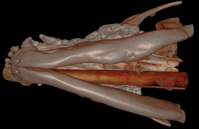
We had the pleasure of seeing Daisy for her four-month follow-up after the ReGum application, and her progress has been remarkable! The bone graft site has healed well, with smooth margins overall, though there are still some minor defects continuing to heal. Importantly, there is no evidence of any further cystic formation.
Daisy’s owners are thrilled with both her appearance and the success of her treatment so far. What’s especially exciting is the amount of bone seen at this stage—significantly more than anticipated, as radiographic mineralization typically takes around six months to become noticeable.
Daisy’s next imaging is scheduled with Animal Dentistry Referral Services for March 2025, and we’re eager to see her continued recovery.
Below: 3D Hard Tissue Overview (left side)
.jpg?width=790&height=505&name=Follow%20Up%20-%203D%20Hard%20Tissue%20Overview%20(left%20side).jpg)
Below: 3D Hard Tissue Overview (ventral aspect)
Below: CT Cross section of the prior disease site, sagittal plane, now with significant healing
%20Case%202%20-%20July%202024/Henry.jpeg)
1 min read
Case Summary: In this case study, we delve into the intricate treatment of a complicated crown fracture in a canine tooth, where the pulp is exposed,...
%20-%20August%202024/3D%20image%20of%20open%20pulp%20chamber.jpg)
1 min read
Case Summary: In this case study, we examine a canine tooth that has been worn down to the pulp, leading to a painful exposure that allows bacteria...
1 min read
Case Summary: Guided Tissue Regeneration (GTR) is a technique used to promote the regeneration of bone and soft tissue, enabling the replacement of...