 Jennifer Mathis, DVM, DAVDC, CVPP
1 min read
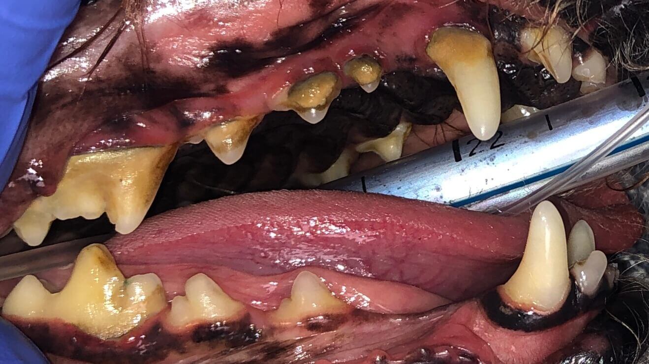
Worn Teeth May Need Root Canal Therapy
Case Summary: In this case study, we examine a canine tooth that has been worn down to the pulp, leading to a painful exposure that allows bacteria...
%20Case%202%20-%20July%202024/Henry.jpeg)
In this case study, we delve into the intricate treatment of a complicated crown fracture in a canine tooth, where the pulp is exposed, causing considerable discomfort for the animal. Such fractures often result from chewing on hard objects like bones, antlers, or hooves. When faced with this issue, veterinarians typically have two options: extraction or Root Canal Therapy (RCT). When deciding on a treatment plan, keep in mind canines are included in the eight important structural teeth, so the best choice is to perform root canal therapy, if possible.
Root Canal Therapy, also referred to as endodontics, entails the meticulous removal of the pulp and thorough cleansing of the tooth's bony walls using specialized files of varying lengths, shapes, and effects. The area is then flushed, disinfected, and dried. Subsequently, the root canal is filled with gutta-percha, a rubber-like material, to effectively seal off the tooth from the rest of the body. Finally, the opening is sealed with tooth-colored composite, akin to the process used by dentists to address cavities in humans.
While the procedure may appear straightforward, it demands a plethora of techniques and requires expensive supplies to be executed with precision and effectiveness.
Henry, a 4-year-old neutered male Labrador Retriever, was referred for evaluation due to a broken lower left canine tooth (304), suspected to have occurred several months prior as a result of an accident.
Below: 3D Overview before procedure: (left and right)
%20Case%202%20-%20July%202024/3D%20Overview.jpg?width=417&height=324&name=3D%20Overview.jpg)
%20Case%202%20-%20July%202024/3D%20Overview%20(1).jpg?width=415&height=327&name=3D%20Overview%20(1).jpg)
Below: Radiograph of CCF - crown, before RCT and restoration
%20Case%202%20-%20July%202024/Radiograph%20of%20ccf%20before%20rct.jpg?width=285&height=219&name=Radiograph%20of%20ccf%20before%20rct.jpg)
%20Case%202%20-%20July%202024/Apex%20radiograph%20before%20RCT.jpg?width=373&height=287&name=Apex%20radiograph%20before%20RCT.jpg)
A root canal procedure was performed on the lower left canine tooth 304, which presented with a complicated crown fracture. To access the pulp chamber, we employed a Piezo opener, ensuring precise and efficient entry.
Below: Images of 304 before RCT and restoration
%20Case%202%20-%20July%202024/complicated%20crown%20fracture%20(ccf)%20(1).jpg?width=373&height=393&name=complicated%20crown%20fracture%20(ccf)%20(1).jpg)
%20(2).jpg?width=423&height=393&name=complicated%20crown%20fracture%20(ccf)%20(2).jpg)
%20Case%202%20-%20July%202024/complicated%20crown%20fracture%20(ccf).jpg?width=373&height=280&name=complicated%20crown%20fracture%20(ccf).jpg)
Upon reaching the pulp chamber, a radiograph was captured to verify cleaning at the designated working length, determined to be 42mm in this case.
Below: Radiograph finding working length
%20Case%202%20-%20July%202024/Working%20length%20radiograph%20during%20RCT.jpg?width=373&height=287&name=Working%20length%20radiograph%20during%20RCT.jpg)
Cleaning and shaping of the canal were meticulously carried out using Dentsply NiTi and LightSpeed files, alternating with suction-assisted sterile saline and sodium hypochlorite solutions to maintain optimal cleanliness.
Below: Radiograph finding master file size to clean canal walls appropriately and find master cone size of gutta percha - note how spade of light speed file fills entire apex
%20Case%202%20-%20July%202024/Master%20file%20during%20RCT%20.jpg?width=373&height=287&name=Master%20file%20during%20RCT%20.jpg)
Following thorough cleaning, a dry fit of gutta-percha was meticulously positioned, and a radiograph was taken to ensure precise placement.
Below: Radiograph dry fitting gutta percha to ensure fills space at apex
%20Case%202%20-%20July%202024/Dry%20fit%20gutta%20percha%20during%20RCT%20procedure%20.jpg?width=373&height=485&name=Dry%20fit%20gutta%20percha%20during%20RCT%20procedure%20.jpg)
Subsequently, the test size was removed, and the canal was meticulously filled with a liquid variant of gutta-percha, ensuring optimal coverage up to the working length. Any excess gutta-percha was trimmed, and the canal walls were meticulously cleaned using specialized techniques.
Below: Radiograph of apex with final fill
%20Case%202%20-%20July%202024/Final%20fill%20apex%20RCT%20rad.jpg?width=373&height=287&name=Final%20fill%20apex%20RCT%20rad.jpg)
Next, a glass ionomer intermediary layer was applied, followed by light-curing and acid-etching to ensure optimal adhesion. An eighth-generation bonding agent (unfilled resin) was then meticulously applied and cured, with additional filled composite layers utilized to effectively seal the access site.
For the final restoration, Omnichroma flowable restoration material was applied and light-cured, ensuring a seamless integration with the surrounding tooth structure. The restoration was then meticulously smoothed to achieve an impeccable finish.
Below: Radiograph of crown with final oil and restorative material
%20Case%202%20-%20July%202024/Final%20fill%20and%20Restoration%20of%20Crown%20RCT.jpg?width=373&height=287&name=Final%20fill%20and%20Restoration%20of%20Crown%20RCT.jpg)
Below: Photographs of 304 after RCT and restorative material
%20Case%202%20-%20July%202024/After%20restoration%20and%20RCT..jpg?width=386&height=275&name=After%20restoration%20and%20RCT..jpg)
%20Case%202%20-%20July%202024/After%20restoration%20and%20RCT.jpg?width=393&height=271&name=After%20restoration%20and%20RCT.jpg)
%20Case%202%20-%20July%202024/Henry%20Ball%204.jpeg?width=386&height=515&name=Henry%20Ball%204.jpeg)
We're thrilled to share that Henry is thriving after his root canal therapy procedure! An update from Henry's person reveals that "He's returned to his lively self at home. Previously, he had been hesitant to engage in his favorite pastime of chasing tennis balls, which is by far his greatest obsession.
However, since his dental procedure, he's been fetching and playing with no signs of discomfort. Henry is exuding happiness once again!"
We're delighted to hear about Henry's progress! Moving forward, he will continue to undergo annual anesthetic dental procedures to ensure the ongoing success of his previous root canal therapy. This is a vital step in maintaining his dental health and overall well-being.
It's worth noting that annual anesthetic dental procedures are recommended for all pets. Studies have demonstrated that pets receiving these regular cleanings live up to 20% longer than those who do not. This underscores the importance of proactive dental care in extending the lifespan and enhancing the quality of life for our furry companions.
.Regular Veterinarian: Cedar Rapids Animal Hospital located at 1000 Memorial Dr SE in Cedar Rapids, IA. We appreciate the referral!
%20-%20August%202024/3D%20image%20of%20open%20pulp%20chamber.jpg)
1 min read
Case Summary: In this case study, we examine a canine tooth that has been worn down to the pulp, leading to a painful exposure that allows bacteria...
1 min read
Case Summary: Guided Tissue Regeneration (GTR) is a technique used to promote the regeneration of bone and soft tissue, enabling the replacement of...
1 min read
Case Summary: Stomatitis is a painful condition characterized by widespread inflammation of the mouth's mucous lining, often linked to immune system...