 Jennifer Mathis, DVM, DAVDC, CVPP
1 min read
Complicated Dental Fractures: The Benefits of Root Canal Therapy
Case Summary: In this case study, we delve into the intricate treatment of a complicated crown fracture in a canine tooth, where the pulp is exposed,...
%20-%20August%202024/3D%20image%20of%20open%20pulp%20chamber.jpg)
In this case study, we examine a canine tooth that has been worn down to the pulp, leading to a painful exposure that allows bacteria to enter and cause disease. Wear patterns, known as attrition, typically develop from repetitive play behaviors, such as chewing on a tennis ball or playing with a frisbee.
In this particular case, you’ll notice that the other three canine teeth have also been worn down, but the body has naturally responded by forming tertiary dentin over the pulp exposure sites. However, in the fourth canine, the wear pattern was more severe, resulting in pulp exposure. To preserve the tooth and maintain its structural integrity, Root Canal Therapy was required.
Root Canal Therapy, or endodontics, involves the removal of the pulp and a thorough cleaning of the tooth’s internal walls using specialized files of varying lengths, shapes, and functions. After the cleaning, the area is flushed, disinfected, and dried. The root canal is then filled with gutta-percha, a rubber-like material that seals the tooth from the rest of the body. Finally, the opening is sealed with a tooth-colored composite, similar to how human cavities are treated.
While the procedure might seem straightforward, it requires a range of techniques and specialized, costly supplies to ensure it is executed effectively.
Condor, a 5-year-old neutered German Shepherd Dog (GSD), was referred for evaluation of worn canines.
Below: 3D Overview Before Procedure: (left and right)
%20-%20August%202024/3D%20Overview%20(1).jpg?width=415&height=334&name=3D%20Overview%20(1).jpg)
%20-%20August%202024/3D%20Overview.jpg?width=368&height=334&name=3D%20Overview.jpg)
Below: 3D Image of Open Pulp Chamber
%20-%20August%202024/3D%20image%20of%20open%20pulp%20chamber.jpg?width=285&height=221&name=3D%20image%20of%20open%20pulp%20chamber.jpg)
%20-%20August%202024/3D%20image%20of%20Attrition%20vs%20open%20pulp%20chamber.png?width=373&height=368&name=3D%20image%20of%20Attrition%20vs%20open%20pulp%20chamber.png)
A root canal procedure was performed on the lower left canine tooth 304, which had a wear pattern that exposed the pulp. A Piezo opener was used to access the pulp chamber. Once access was gained, a radiograph was taken to confirm the working length, which measured 45mm in this case.
Below: Worn Canine with Open Pulp Chamber - (Image of Pathfinder)
%20-%20August%202024/Worn%20canine%20with%20open%20pulp%20chamber%20-%20image%20of%20pathfinder%20.jpg?width=263&height=366&name=Worn%20canine%20with%20open%20pulp%20chamber%20-%20image%20of%20pathfinder%20.jpg)
Below: Radiograph Showing Working Length
%20-%20August%202024/Radiograph%20showing%20working%20length%20during%20RCT.jpg?width=373&height=287&name=Radiograph%20showing%20working%20length%20during%20RCT.jpg)
Below: Radiograph Showing Master File Size
%20-%20August%202024/Radiograph%20showing%20master%20file%20size%20during%20RCT.jpg?width=373&height=287&name=Radiograph%20showing%20master%20file%20size%20during%20RCT.jpg)
The canal was then cleaned and shaped using Dentsply NiTi and LightSpeed files, alternating with suction-assisted sterile saline and sodium hypochlorite solutions. After thorough cleaning, a dry fit of gutta-percha was placed, followed by a radiograph to verify the fit. The gutta-percha was then removed, and the canal was filled with a liquid version of gutta-percha. A solid gutta-percha point was pressed to the working length, trimmed to remove excess, and the canal walls were cleaned using specialized techniques.
Below: Radiograph of Dry Fit of Gutta-Percha
%20-%20August%202024/Radiograph%20of%20dry%20fit%20of%20gutta%20percha%20during%20RCT.jpg?width=374&height=287&name=Radiograph%20of%20dry%20fit%20of%20gutta%20percha%20during%20RCT.jpg)
A glass ionomer intermediary layer was applied, light-cured, and acid-etched. An eighth-generation bonding agent (unfilled resin) was then applied and cured, followed by additional layers of filled composite to seal the access site. For the final restoration, iM3 Create packable and Omnichroma flowable materials were used, light-cured, and smoothed for a polished finish.
Below:Radiograph of Final Fill of RCT - Apex
%20-%20August%202024/Radiograph%20of%20Final%20fill%20of%20RCT%20-%20Apex.jpg?width=373&height=287&name=Radiograph%20of%20Final%20fill%20of%20RCT%20-%20Apex.jpg)
Below: Radiograph of Final Restoration of RCT - Crown
%20-%20August%202024/Radiograph%20of%20Final%20restoration%20of%20RCT%20-%20Crown%20.jpg?width=373&height=287&name=Radiograph%20of%20Final%20restoration%20of%20RCT%20-%20Crown%20.jpg)
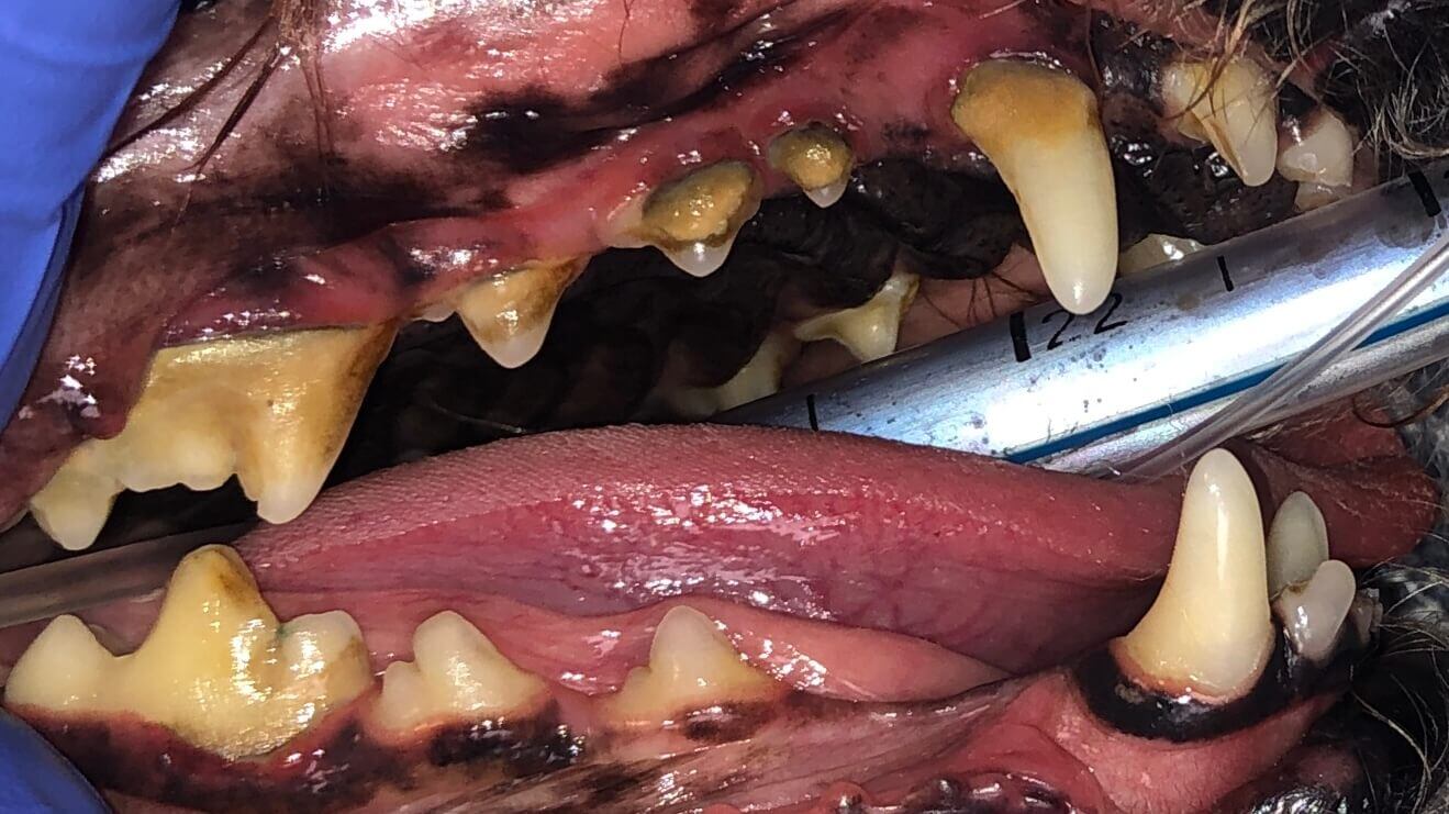
Below: Side Views Before Dental Cleaning with Worn Canine (Attrition and Tertiary Dentin over 404)
%20-%20August%202024/Side%20view%20before%20RCT%20and%20dental%20cleaning.jpg?width=386&height=214&name=Side%20view%20before%20RCT%20and%20dental%20cleaning.jpg)
%20-%20August%202024/Side%20view%20before%20dental%20cleaning%20with%20worn%20canine%20-%20Attrition%20and%20Tertiary%20Dentin%20over%20404.jpg?width=433&height=214&name=Side%20view%20before%20dental%20cleaning%20with%20worn%20canine%20-%20Attrition%20and%20Tertiary%20Dentin%20over%20404.jpg)
Below: Side Views After Dental Cleaning
%20-%20August%202024/Side%20view%20-%20After%20RCT.jpg?width=424&height=214&name=Side%20view%20-%20After%20RCT.jpg)
%20-%20August%202024/Side%20view%20after%20dental%20cleaning%20.jpg?width=351&height=214&name=Side%20view%20after%20dental%20cleaning%20.jpg)
Below: Top View Before (left) and After (right) RCT Restoration
%20-%20August%202024/Top%20view%20before%20procedure.png?width=258&height=366&name=Top%20view%20before%20procedure.png)
%20-%20August%202024/Top%20view%20of%20restoration%20after%20RCT.jpg?width=383&height=366&name=Top%20view%20of%20restoration%20after%20RCT.jpg)
Condor is doing great at home after his root canal therapy! His recovery from anesthesia was smooth, and he's back to enjoying his favorite activities without any discomfort. To prevent future tooth fractures, it's important to ensure that all pets only chew on flexible items for life—no bones, hooves, nylabones, antlers, or non-flexible rawhides.
Interestingly, a study involving 2 million dogs found that those receiving annual anesthetic dental procedures lived 20% longer. Regular dental care is key to a healthy and happy life!
%20Case%202%20-%20July%202024/Henry.jpeg)
1 min read
Case Summary: In this case study, we delve into the intricate treatment of a complicated crown fracture in a canine tooth, where the pulp is exposed,...
1 min read
Case Summary: Guided Tissue Regeneration (GTR) is a technique used to promote the regeneration of bone and soft tissue, enabling the replacement of...
1 min read
Case Summary: Stomatitis is a painful condition characterized by widespread inflammation of the mouth's mucous lining, often linked to immune system...