 Jennifer Mathis, DVM, DAVDC, CVPP
1 min read
Complicated Dental Fractures: The Benefits of Root Canal Therapy
Case Summary: In this case study, we delve into the intricate treatment of a complicated crown fracture in a canine tooth, where the pulp is exposed,...
%20-%20March%202024/current%20pic.jpg)
Most discolored (non-vital) teeth are essentially dead, their discoloration resulting from the breakdown of pulp contents leaching into the dentinal tubules within the tooth. This process is not merely cosmetic; it's also quite painful. Moreover, these breakdown products create an ideal environment for bacterial colonization, worsening the issue. The only viable options for treatment are root canal therapy or extraction.
Root canal therapy, also known as endodontics, involves the removal of the pulp and thorough cleaning of the tooth's bony walls using specialized files of different lengths, shapes, and effects. The area is then flushed, disinfected, and dried. Subsequently, the root canal is filled with gutta-percha, a material similar to rubber, to seal the tooth off from the rest of the body. Finally, the opening is sealed with tooth-colored composite, akin to how a dentist seals human cavities.
While the procedure may seem straightforward, it requires numerous techniques and expensive supplies to be executed effectively.
Tucker, a 5-year-old male neutered American Pitbull Terrier, was seen for a discolored upper left canine (204). This discoloration had been present for many months. The pet owner was previously advised that extraction was the only treatment option, and declined treatment. The tooth was left non-vital until self-referral to Animal Dentistry Referral Services for Root Canal Therapy (RCT) in October of 2021.
CT Imaging showed:
T/NV (non vital tooth) 204.
Missing 408 and 311, 411.
Beginning RR (root resorption) to lower rostral premolars - a common finding for dogs of this age and size
Below: 3D Overview before procedure: (left and right)
%20-%20March%202024/3D%20Overview.jpg?width=417&height=374&name=3D%20Overview.jpg)
%20-%20March%202024/3D%20Overview%20(1).jpg?width=415&height=375&name=3D%20Overview%20(1).jpg)
Below: Discolored tooth on CT Imaging
%20-%20March%202024/CT%20Image%20of%20discolored%20204.jpg?width=285&height=412&name=CT%20Image%20of%20discolored%20204.jpg)
%20-%20March%202024/CT%20Image%20comparing%20upper%20canines%20root%20canals%20104%20vs%20discolored%20204.jpg?width=373&height=281&name=CT%20Image%20comparing%20upper%20canines%20root%20canals%20104%20vs%20discolored%20204.jpg)
Below: Radiograph of 204 of the apex (tip of root). Beyond the wide pulp chamber, nothing significant is noted, yet studies show 43% of discolored teeth have no radiographic signs of problems, but have pulp necrosis and disease that leads to additional, potentially systemic issues.
%20-%20March%202024/Radiograph%20before%20RCT%20-%20apex.jpg?width=386&height=297&name=Radiograph%20before%20RCT%20-%20apex.jpg)
Below: Before RCT - Radiograph of 204 to aid in planning the access point
%20-%20March%202024/radiograph%20before%20RCT%20-%20access%20point.jpg?width=386&height=297&name=radiograph%20before%20RCT%20-%20access%20point.jpg)
A root canal procedure was performed on tooth 204, which was non-vital. To access the pulp chamber, a round surgical length bur was utilized. Upon reaching the pulp chamber, a radiograph was taken to ensure cleaning at the working length, which in this case was 38mm. Cleaning and shaping of the canal were carried out using Dentsply NiTi and LightSpeed files in alternation with suction aided sterile saline and sodium hypochlorite solutions.
Below: During RCT - Radiograph finding working length to clean and measure canal
%20-%20March%202024/during%20RCT%20-%20finding%20working%20length.jpg?width=379&height=291&name=during%20RCT%20-%20finding%20working%20length.jpg)
Below: During RCT - finding master file size to clean canal walls appropriately and find master cone size of gutta percha
%20-%20March%202024/Radiograph%20of%20master%20file%20during%20RCT.jpg?width=386&height=296&name=Radiograph%20of%20master%20file%20during%20RCT.jpg)
Below: After cleaning, a gutta-percha point was sized to aid in filling, with radiographic confirmation of reaching the tip.
%20-%20March%202024/Radiograph%20of%20dry%20fit%20of%20gutta%20percha%20during%20RCT.jpg?width=386&height=297&name=Radiograph%20of%20dry%20fit%20of%20gutta%20percha%20during%20RCT.jpg)
The test size was then removed, and the canal was filled with a liquid version of gutta percha, pressing in the solid gutta-percha point to the working length. Excess gutta-percha was trimmed and the walls cleaned using special techniques.
Below: After RCT - final mid root fill of solid and liquid turned solid gutta percha.
%20-%20March%202024/Radiograph%20of%20final%20fill%20of%20RCT%20-%20middle.jpg?width=386&height=297&name=Radiograph%20of%20final%20fill%20of%20RCT%20-%20middle.jpg)
A glass ionomer intermediary layer was applied. This was light-cured and acid-etched, then a eighth generation bonding agent (unfilled resin) was applied and cured with additional filled composite layers applied to seal the access site.
Below: After RCT - final restoration with radiolucent unfilled resin layer.
%20-%20March%202024/Radiograph%20of%20final%20fill%20of%20RCT%20-%20crown.jpg?width=386&height=297&name=Radiograph%20of%20final%20fill%20of%20RCT%20-%20crown.jpg)
Packable composite was used for the final restoration, as a crown was not utilized, as additional strength was deemed beneficial. Light curing was performed in 2mm increments, and special sanding and polishing discs were used for smoothing the final restoration.
Below: Overview before procedure: (left and right)
%20-%20March%202024/Overview%20of%20left%20side%20before%20RCT%20and%20cleaning.jpg?width=386&height=245&name=Overview%20of%20left%20side%20before%20RCT%20and%20cleaning.jpg)
%20-%20March%202024/Overview%20of%20right%20side%20before%20dental%20cleaning.jpg?width=386&height=240&name=Overview%20of%20right%20side%20before%20dental%20cleaning.jpg)
Below: Overview after procedure: (left and right)
%20-%20March%202024/Overview%20of%20left%20side%20after%20RCT%20and%20cleaning.jpg?width=386&height=213&name=Overview%20of%20left%20side%20after%20RCT%20and%20cleaning.jpg) .
. %20-%20March%202024/Overview%20of%20right%20side%20after%20dental%20cleaning.jpg?width=386&height=219&name=Overview%20of%20right%20side%20after%20dental%20cleaning.jpg)
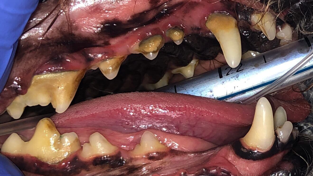
Below: (left) Close up of non-vital 204 before RCT (right) Close up of non-vital 204 after RCT
*Note that performing root canal therapy cannot remove all of the staining located in the dentinal tubules.%20-%20March%202024/Image%20of%20204%20before%20RCT.jpg?width=386&height=578&name=Image%20of%20204%20before%20RCT.jpg)
%20-%20March%202024/Image%20of%20204%20after%20RCT.jpg?width=386&height=418&name=Image%20of%20204%20after%20RCT.jpg)
%20-%20March%202024/current%20pic.jpg?width=386&height=510&name=current%20pic.jpg)
Tucker is thriving at home! At 7 years old, he hasn't slowed down one bit. Being a beloved pet of the ADRS staff, we get to see him frequently. Although his tooth will always remain discolored, he is pain-free and undergoes annual anesthetic dental procedures to ensure there are no issues with his previous root canal therapy.
It's recommended that every pet undergoes an annual anesthetic dental procedure, as studies have shown that pets receiving these annual anesthetic cleanings live 20% longer than those who do not.
Regular Veterinarian/s: Dr. Jennifer Mathis and Dr. Ryan Southard at Family Pet Veterinary Center.
%20Case%202%20-%20July%202024/Henry.jpeg)
1 min read
Case Summary: In this case study, we delve into the intricate treatment of a complicated crown fracture in a canine tooth, where the pulp is exposed,...
%20-%20August%202024/3D%20image%20of%20open%20pulp%20chamber.jpg)
1 min read
Case Summary: In this case study, we examine a canine tooth that has been worn down to the pulp, leading to a painful exposure that allows bacteria...
1 min read
Case Summary: Guided Tissue Regeneration (GTR) is a technique used to promote the regeneration of bone and soft tissue, enabling the replacement of...