1 min read
Treating Periodontal Disease: Ziggy's Journey with Guided Tissue Regeneration
Case Summary: Guided Tissue Regeneration (GTR) is a technique used to promote the regeneration of bone and soft tissue, enabling the replacement of...
2 min read
 Jennifer Mathis, DVM, DAVDC, CVPP
:
May 1, 2024 12:00:00 AM
%20-%20February%202024/Canine%20Chronic%20Ulcerative%20Stomatitis%20(CCUS)-2.png)
Canine Chronic Ulcerative Stomatitis (CCUS) is characterized by painful ulcerative lesions in the mucosal epithelium, believed to result from an immune response to plaque on the teeth. These ulcerative lesions may develop in areas lacking teeth as well as in paradental regions, possibly extending to the tongue. CCUS is a permanent and long term condition. Medications alone historically prove insufficient for managing the disorder.
In this case study, the patient underwent his routine anesthetic dental procedure with his regular veterinarian, Dr. Ryan Southard, from Family Pet Veterinary Center, during which this condition was identified.
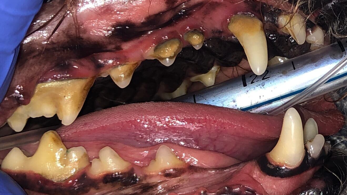
A 2-year-old male neutered (MN) Boxer, exhibited initial exam findings including a calculus index (CI) of 3 and a gingival index (GI) of 3, with marked gingival recession, bleeding, and purulent exudate overlying bilateral upper carnassials.
He presented for comprehensive oral health assessment and treatment (COHAT) two months after the initial examination, during which the referring veterinarian diagnosed Canine Chronic Ulcerative Stomatitis (CCUS).
"Kissing lesions" (ulcerative lesions) were noted under anesthesia by the referring veterinarian (RDVM), who also reported "brown drool" on the day of the dental procedure. Mobile incisor teeth and a lower right premolar were extracted by the RDVM, and the patient was subsequently referred to Animal Dentistry Referral Services for further care.

Missing teeth: 408, 307.
Previously extracted: 101, 201, 407, 303 with referring DVM.
FE3 (Furcation exposure through and through): 108, 208, severe ulcerative lesions in tissues adjacent to 108, 208.
Severe marginal inflammation, worse over 300 quadrant.
Below: Hard tissue 3D reconstruction using CBCT

Below: (left) Hard tissue 3D CBCT - front, (right) Wear on lower canines due to underbite

Below: Bone loss (FE3 upper fourth premolars) - need treatment
%20-%20need%20treatment.png?width=2078&height=624&name=Bone%20loss%20(FE3%20upper%20fourth%20premolars)%20-%20need%20treatment.png)
CCUS is an awful chronic disease. Selective tooth extraction along with medical management is the treatment of choice.
The teeth were scaled to remove plaque buildup. SANOS application was administered to reduce plaque accumulation, while doxycycline was prescribed to downregulate matrix metalloproteinases responsible for soft tissue destruction. Daily use of MaxiGuard wipes, containing Zinc, aimed to further reduce plaque accumulation and aid in gingival tissue health. A follow-up appointment in three months was scheduled to assess the need for additional extractions and monitor CCUS status.
Gingivoplasty 309, 409: Excess or reactive gum tissue was removed during the procedure.
Below: Images showing "Kissing" lesions


Below: Images showing "Kissing" lesions on the tongue


Surgical Extraction 108, 208: Extracted 108, 208. The procedure involved creating a flap, sectioning the teeth, elevating them, performing alveoloplasty, and closing the surgical site with 4-0 monocryl sutures.
Below: Before cleaning

Below: After cleaning & before extractions

Below: (left) Before extractions, (right) After extractions

Below: Before extraction highlighting sites of gingival contouring

Below: After extractions

The owners reported the patient showed some improvement within the month following the 2021 dental procedure. In March of 2022, about 3 months post procedure, the patient presented with discolored, excessive salivation although owner brushed his teeth daily with MaxiGuard wipes.
Oral findings: Moderate erythema bilaterally on buccal cheeks and lateral tongue margins as well as thickened saliva/mucous; L side worse than R side, dental calculus noted to the upper molars. Owner had restarted Doxycycline 2 days prior (was not directed to stop previously), and the patient referred back to Animal Dentistry Referral Services.
Dental exam findings under anesthesia: Severe contact mucositis, need to perform caudal mouth extraction (CME - all teeth behind canines required extractions) Scaled and polished, but extractions were declined by the owner. As inflammation had returned within 3 months (somewhat less than November 2021), the owner was instructed to schedule CME ASAP - but within 3 moths.
Below: 3D Overview

Below: Needs Caudal Mouth Extraction
%20-%20February%202024/last%20procedure%20need%20CME%20right.png?width=453&height=287&name=last%20procedure%20need%20CME%20right.png)
%20-%20February%202024/last%20procedure%20need%20CME%20left.png?width=452&height=303&name=last%20procedure%20need%20CME%20left.png)
Last update: The patient was still experiencing discolored, excessive salivation and gingival inflammation. Unfortunately the owner cancelled the follow up dental procedure, and has since moved away.
The images show follow up when not treated fully. There is significant localized improvement due to tooth extractions, so it would be expected to be able to be almost fully controlled in this patient had CME been performed.
More resources: There is a recent paper about adjunctive medical management of CCUS by Dr. Kim Ford that can be another tool for CCUS treatment.

1 min read
Case Summary: Guided Tissue Regeneration (GTR) is a technique used to promote the regeneration of bone and soft tissue, enabling the replacement of...
%20-%20March%202024/current%20pic.jpg)
1 min read
Case Summary: Most discolored (non-vital) teeth are essentially dead, their discoloration resulting from the breakdown of pulp contents leaching into...
%20Case%202%20-%20July%202024/Henry.jpeg)
1 min read
Case Summary: In this case study, we delve into the intricate treatment of a complicated crown fracture in a canine tooth, where the pulp is exposed,...