---
title: Western Veterinary Conference 2024
---
[Skip to the main content.](https://tooth.vet/wvc-extlab#main-content)
[/Animal_Dentistry__logo-removebg.png?width=1280&height=720&name=Animal_Dentistry__logo-removebg.png "Animal-Dentistrylogo-transparent")](https://tooth.vet/?hsLang=en)
[/Animal-Dentistrylogo-transparent.png?width=1280&height=720&name=Animal-Dentistrylogo-transparent.png "Animal-Dentistrylogo-transparent")](https://tooth.vet/?hsLang=en)
- [Home](https://tooth.vet)
- [About Us](https://tooth.vet/about-us)
- [Contact Us!](https://tooth.vet/contact-us)
- Services
- [3D Imaging](https://tooth.vet/3d-imaging)
- [In-House Anesthesia](https://tooth.vet/in-house-anesthesiologist)
- [Canine Dentistry](https://tooth.vet/canine-dentistry)
- [Feline Dentistry](https://tooth.vet/feline-dentistry)
- [Dental Conditions](https://tooth.vet/dental-conditions-full-list)
- [Dental Education](https://tooth.vet/dental-education)
- [Patient Referral](https://tooth.vet/patient-referral)
- [Pet Owners](https://tooth.vet/pet-owners)
- [Dentistry Case Chronicles](https://tooth.vet/beneath-the-surface-animal-dentistry-referral-services-case-chronicles)
- [Upcoming Events](https://tooth.vet/events)
Search
Toggle Menu
Search
Toggle Menu
- [Home](https://tooth.vet)
- [About Us](https://tooth.vet/about-us)
- [Contact Us!](https://tooth.vet/contact-us)
- Services
- [3D Imaging](https://tooth.vet/3d-imaging)
- [In-House Anesthesia](https://tooth.vet/in-house-anesthesiologist)
- [Canine Dentistry](https://tooth.vet/canine-dentistry)
- [Feline Dentistry](https://tooth.vet/feline-dentistry)
- [Dental Conditions](https://tooth.vet/dental-conditions-full-list)
- [Dental Education](https://tooth.vet/dental-education)
- [Patient Referral](https://tooth.vet/patient-referral)
- [Pet Owners](https://tooth.vet/pet-owners)
- [Dentistry Case Chronicles](https://tooth.vet/beneath-the-surface-animal-dentistry-referral-services-case-chronicles)
- [Upcoming Events](https://tooth.vet/events)
[Facebook](https://tooth.vet/wvc-extlab#0) [Instagram](https://tooth.vet/wvc-extlab#0) [Linkedin](https://tooth.vet/wvc-extlab#0) [X](https://tooth.vet/wvc-extlab#0) [YouTube](https://tooth.vet/wvc-extlab#0) [Medium](https://tooth.vet/wvc-extlab#0)
## **References and Resources for: Canine & Feline Dental Extraction Lab**
# Extraction Lab
---
### 2025 Lab Downloads
**[Lab Proceedings ](https://23427187.fs1.hubspotusercontent-na1.net/hubfs/23427187/WVC%20Resources/WVC%202025/Jen%20proceedings/Extraction%20lab%20handout%20WVC%202025.pages)**
**[Instrument List ](https://23427187.fs1.hubspotusercontent-na1.net/hubfs/23427187/WVC%20Resources/WVC%202025/Jen%20proceedings/Basic%20instrument%20and%20description%20of%20use.pdf)**
[Download Dental Kit Brochure 👇](https://23427187.fs1.hubspotusercontent-na1.net/hubfs/23427187/WVC%20Resources/WVC%202025/Dr%20Mathis%20new%20dental%20kit%202025%20Updated.pdf)
[.jpeg?width=200&height=200&name=mediamodifier_image%20(20).jpeg "Dr jen tool kit graphic")](https://23427187.fs1.hubspotusercontent-na1.net/hubfs/23427187/WVC%20Resources/WVC%202025/Dr%20Mathis%20new%20dental%20kit%202025%20Updated.pdf)
[mailto:ce@tooth.vet](mailto:ce@tooth.vet)
### 2025 Videos
---
### 2024 Lab Downloads
** [Lab Proceedings](https://23427187.fs1.hubspotusercontent-na1.net/hubfs/23427187/WVC%20Resources/Extraction%20lab%20handout.pdf)**

---

---
We're excited to offer a carefully selected collection of Dentistry focused CE courses designed specifically for DVMs and Technicians. Expand your knowledge and advance your career with our comprehensive library of resources.
### Vet CE You'll Use!
### Guide to Dental Conditions 12-Page Printable E-Book
Gain a wealth of knowledge on over 20 common dental conditions that can impact your veterinary patients. This 12-page printable e-book features detailed descriptions and photos of each condition, along with common treatment approaches.
[Get Your Free Copy Here](https://tooth.vet/wvc-extlab#tmp1_module_17047447312745)
.jpeg)
#### [DOWNLOAD YOUR COPY](https://hubs.ly/Q01K1TD60)
Explore this comprehensive list of over 20 common dental conditions that affect veterinary patients. Simply click on each condition to learn more about them and their potential impact on your pet's oral health.
- #### Persistent Baby Teeth - What You Need to Know 🐕🦺

We all know that puppies and kittens can be a handful. They initially need monthly check-ins with the vet. A series of vaccinations is needed. Hours and hours of puppy training should also be invested. But did you know that growing puppies and kittens also need to have their mouths checked regularly while developing? If you didn't then you're probably asking "Why?" now. Well in this article, we'll talk about everything you need to know about your pet's baby teeth, and how to catch unwanted problems at the "root"!
##### When Do My Pet's Teeth Start Coming In?
Deciduous, aka ‘baby’ teeth, begin to erupt at 3-4 weeks of age with dogs having 28 baby teeth and cats having 26. The adult teeth begin erupting as soon as 3 months of age finishing as late as 7 months of age. A normal adult dog has 42 teeth. A typical cat has 30 adult teeth by 6 months of age.
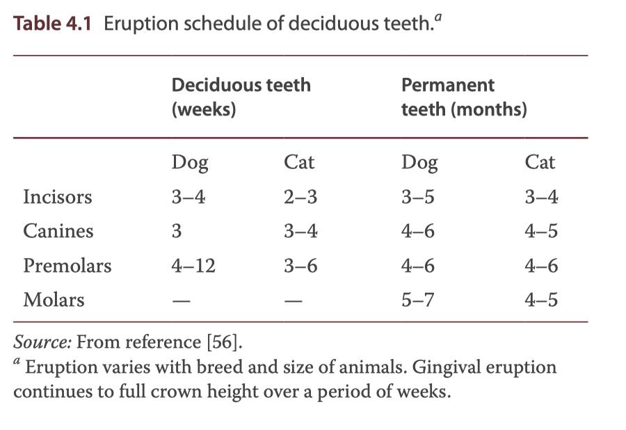
Most of this change happens in the 4-6 month age time period when one can see both adult and baby teeth, known as mixed dentition. Some people say this time can cause your pet to feel irritable or not themselves as teething can be an uncomfortable process. One often sees puppies chewing on things they shouldn’t, or in rare instances, a lack of interest in food. It’s always a good idea to get your pup checked out if you’re noticing these issues, as they could also be linked to other issues going on. If you would like to learn more about teething in puppies and the proper chew toys to use while they're going through this, go to our blog titled "What to Look For in a Chew Toy" or click [here](https://www.fampetvet.com/services/blog/what-look-chew-toy). Remember many things at the store can break teeth and are not appropriate.
They are born with adult teeth present in bone still continuing to form roots before they emerge from the gums. While they’re growing, they begin to put pressure onto the current deciduous teeth and stimulate them to begin reabsorbing. After the baby teeth’s roots have been reabsorbed, the crowns and hollow shells of the teeth are pushed out of the tooth socket and from the mouth. This is what should happen as your pet's adult teeth begin to come in, but it doesn't always work out that way. Many owners also think they will find their dog’s baby teeth. Most of the time, they are swallowed or lost, never to be seen usually without consequence.
##### What Are Persistent Deciduous Teeth?
Baby teeth (also called primary or deciduous teeth) that are still present when the adult teeth begin to emerge are called persistent deciduous teeth. The persistent deciduous teeth cause the forming adult teeth to emerge from the gums in an altered position.
Persistent deciduous teeth could lead to more secondary issues within your pet's mouth. Not only will the adult teeth and baby teeth be in the same spot, but the main concerns are secondary alignment issues causing a painful bite and periodontal disease due to crowding. With more teeth in the mouth, it is more likely for food, hair, and other debris to collect around teeth and cause inflammation. This can create faster periodontal disease development, which is commonly seen as the red swelling in your pet's mouth and can have degrees of bone loss below the gum line. The rule to remember is that two teeth should never be in the same spot at the same time after six months of age. Sometimes we need to intervene earlier as well. it is important to remove the baby tooth as soon as the problem arises especially in small dog breeds that are more likely to develop both persistent baby teeth AND periodontal disease.
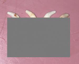
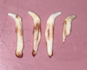
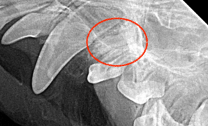
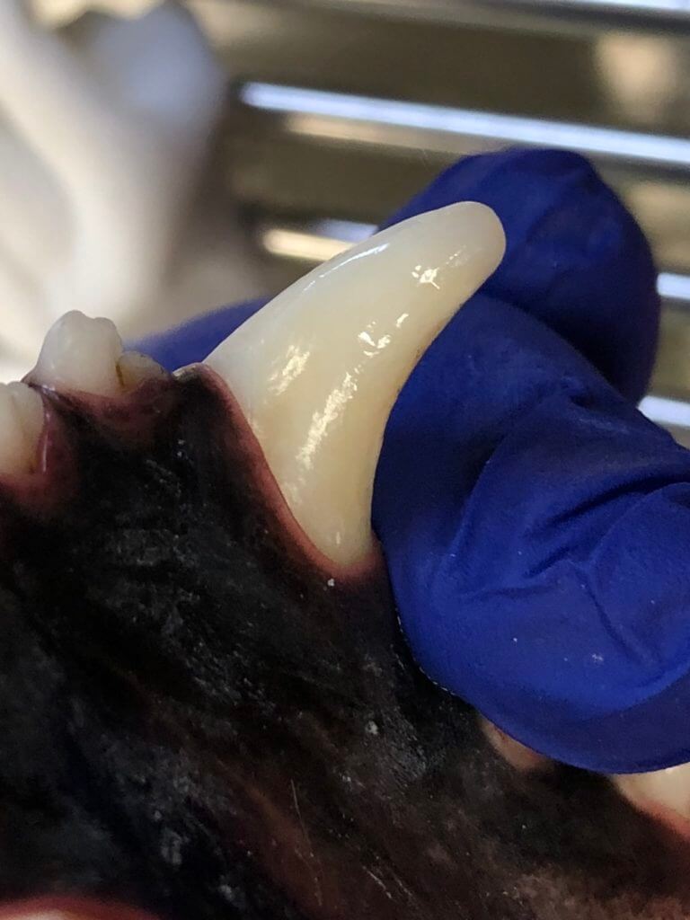
Removing baby teeth is not as simple as one would think. The roots are often 3 to 4 times as long as the crown. The first photo shows the tooth, the second shows the full tooth including root.
|  |  |
| --- | --- |
If roots happen to break, they have the risk of causing eye and nasal issues. There is a common conception that broken roots will be resorbed by the body, but this happens much less frequently than veterinarians as a whole think it does. The retained tooth root contains the pulp (blood vessels and nerves), a source of pain and disease. These become a chronic source of hidden pain that your pet cannot tell you about. This is just one reason for the importance of taking full mouth intraoral x-rays on every anesthetic dental procedure.
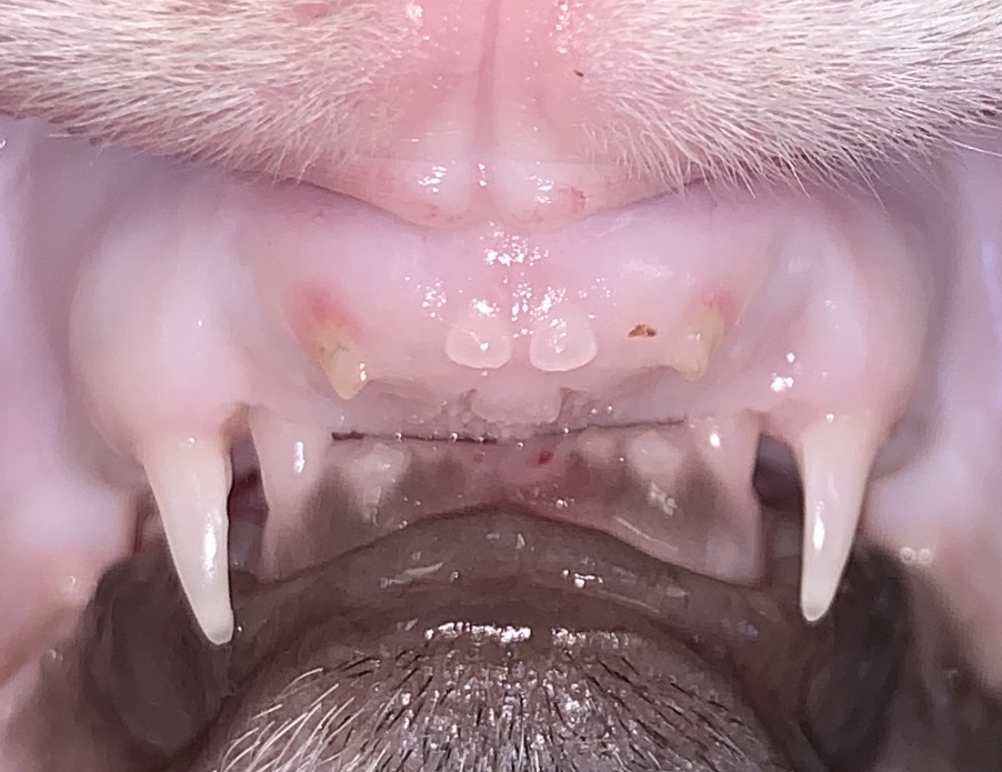
Another common problem seen with persistent deciduous teeth would be if it was a lower canine that was affected: In this case, the adult tooth erupts too close on the tongue side because the baby teeth has not been lost (as mentioned above). This results in the lower canines poking into the roof of the mouth and causing open wounds, infection, or even upper tooth or nose damage. Often times this is accompanied by the lower mandibles being too short, a growth discrepancy that is usually genetically predisposed. With the mandibles being too short, there isn’t enough space for the canines to tip outward and interdigitate with the upper teeth. Sometimes a baby tooth may not yet be persistent but the jaw length relationship allows the lower canines to catch in the soft tissues of the palate preventing the appropriate growth.
##### Is My Pet At Risk for Persistent Deciduous Baby Teeth?
Small dogs or dog breeds are more likely to develop persistent deciduous teeth and especially brachycephalic (smush-faced) dogs. Any baby tooth could end up being persistent, but it’s most commonly in the upper canines. After that would be the lower canines and then the incisors.
If your puppy has a jaw length discrepancy or the lower canines (baby and/or adult) erupt to contact the palate with or without persistent deciduous teeth, this condition can be hereditary. It is not recommended to breed dogs with this condition even if it is corrected.
##### Are There Treatment Options for Persistent Deciduous Baby Teeth?
Yes! Persistent baby teeth should be extracted as soon as they are noticed. It will require general anesthesia and careful precautions will be taken by the performing veterinarian to try to prevent the crowns of the adult teeth to be damaged during the procedure. If the persistent baby teeth are not noticed until neutering, then it is okay to have it done during the same procedure. Waiting to have the baby teeth removed could lead to malposition of the adult tooth, slowing the growth of the lower jaw, and/or bone loss around the adult tooth. The sooner the problem is found and treated, is more likely the resolution to be faster and simpler.
After discharge from the dental extraction, your pet should remain on a soft diet for 10-14 days after the dental procedure. This means wet food or if your pet won’t eat wet food then dry kibble softened and soaked in water. In addition to using pain management during the dental procedure, your veterinarian will usually send you home with additional pain medication for 3-5 days after surgery. Brushing and wipes should be avoided until 10-14 days after the procedure when your pet comes back in for the Medical Progress Exam. This re-evaluation appointment is to ensure that your pet is healing properly from the dental procedure and no additional problems have arisen.
A persistent baby tooth without an adult counterpart seen visually or radiographically (on x-ray), is an exception. It can be treated like any normal tooth. It usually has thinner, longer roots, but can stay as long as it is healthy on the margin and inside as we evaluate all teeth.
##### Is There Any Kind of Prevention?
There is no prevention as the main influence is genetics. The best way to avoid any serious complications would be to catch the situation as early as possible. This is part of the evaluation your veterinarian should look at during the puppy visits which are typically at 6, 9, 12, and 16 weeks of age. Having additional visits at 20 and 24 weeks can be helpful to catch changes during this rapid growth phase. You can also begin brushing your pet's teeth and cleaning with dental wipes at 2 weeks of age, but you should remain gentle during this time as the teeth coming in are fresh and tender. Brushing or wipes should be used daily or at least every other day to be effective in dental care. If you keep this habit up and start early, it’s much easier to continue as they grow older. Additionally, by brushing your pet’s teeth every day, you’re able to get a good look at the mouth and notice any differences or issues as soon as they arise.
##### Final Verdict:
Two teeth in the same location are a serious concern that should be evaluated as soon as it is discovered. Additionally, the earlier the problem is found, one can have easier methods to correct it before the problem worsens. If you notice anything that doesn’t look right or any additional teeth where they shouldn’t be, give us a call or schedule an appointment with us to get it checked out!
- #### Passive Force Orthodontics for Base Narrow Canines 🐕🦺
Dental conditions are often hidden and painful. The following posts are going to help describe conditions that pets can get as well as treatment. Many people I talk with are surprised at how we can help pets with dental conditions and save teeth whenever possible.
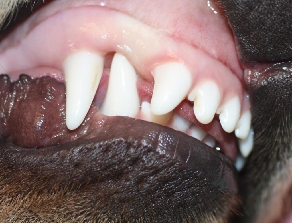
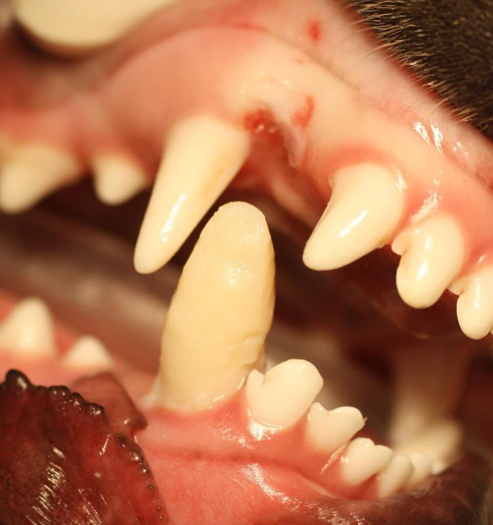
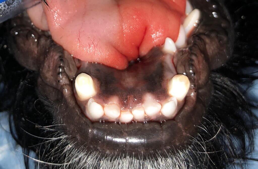
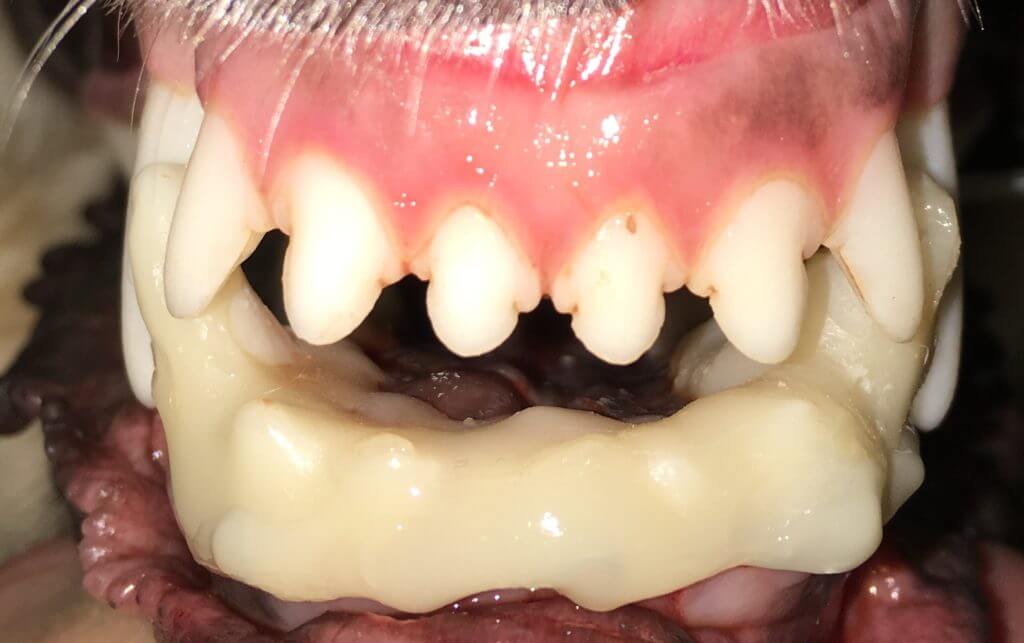
Sometimes we see baby lower canine teeth located too far towards the middle of the lower jaw. These sharp needle-like teeth often poke into the roof of the mouth delaying appropriate jaw growth. Any time the lower canines (adult or baby) contact the roof of the mouth, this needs to be corrected. We can use temporary acrylic appliances molded to the teeth to help the teeth to move
A common problem often found late in the course of progression is linguoverted canines. This means the lower canine teeth (aka “fangs”) are erupting nearer the tongue and contacting the roof of the mouth.
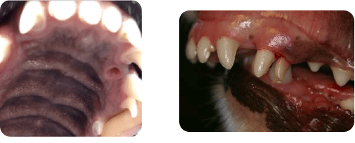
This is another of the hidden painful conditions. If allowed to progress, two common sequelae occur a hole in the roof of the mouth (not good) and/or the lower jaw getting stuck in place and cannot grow appropriately during puppyhood resulting in misalignment that can have pain and crowding progressing to infection. Skeletal maturity is reached often near 10 months of age in dogs making the jawless likely to continue to grow. Treatments can still be done but none will result in the correction of all alignment. The sooner this problem is found and treated, the more likely the body will correct itself.
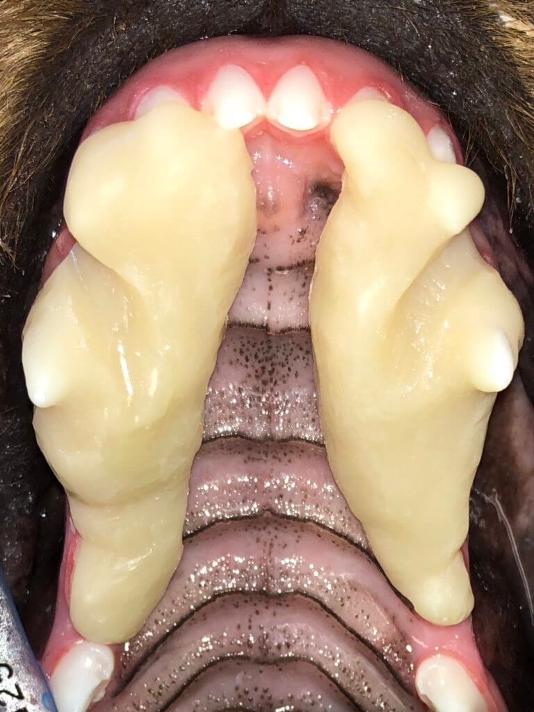
Sometimes we use incline planes to change the angle of the tooth naturally as a dog bites down. This is usually placed for 3-6 weeks. After the acrylic is removed the surrounding soft tissues are irritated but heal up in just a few days.
Other times we are fortunate to be able to just use acrylic composites to extend the length of the canine tooth, helping the tooth to tip into the correct position.
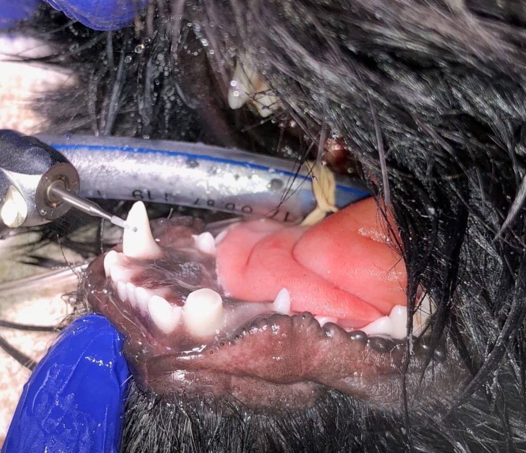
If a patient will not tolerate one of the above treatments or is an anesthetic risk requiring a single procedure, an intentional crown shortening can be performed with vital pulp therapy (miniature root canal therapy) to save the shortened tooth. It is always better and usually more successful to keep the full normal tooth structure when possible, thus vital pulp therapy; is not a first-choice treatment.
|  |  |
| --- | --- |
|  |  |
| --- | --- |
Occasionally this condition is mild and will be caught very early thus is corrected with only extraction of the baby teeth and recontouring of the gumline:
| 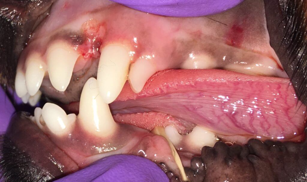 | 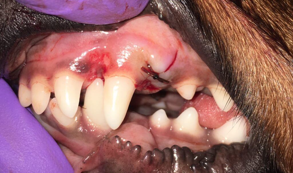 |
| --- | --- |
This is usually a genetic condition and breeding a pet after one of these procedures is unethical due to the risk of passing this condition on to offspring.
- #### Abnormal bite (occlusion) 🐕🦺
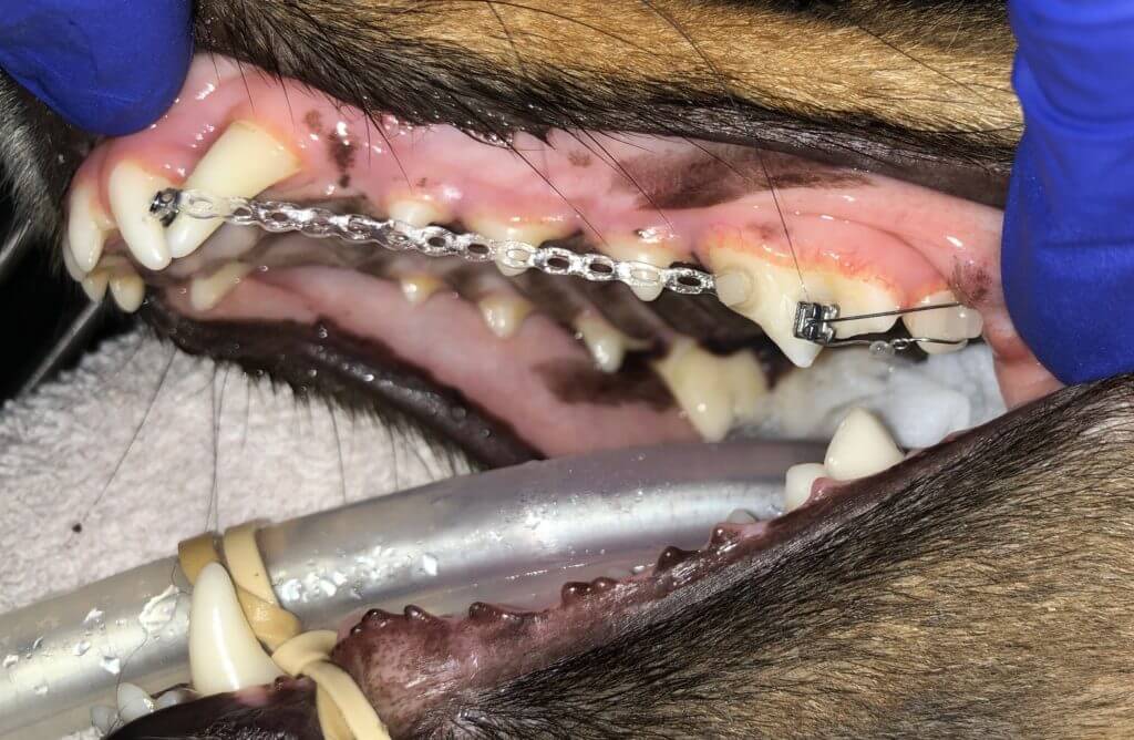
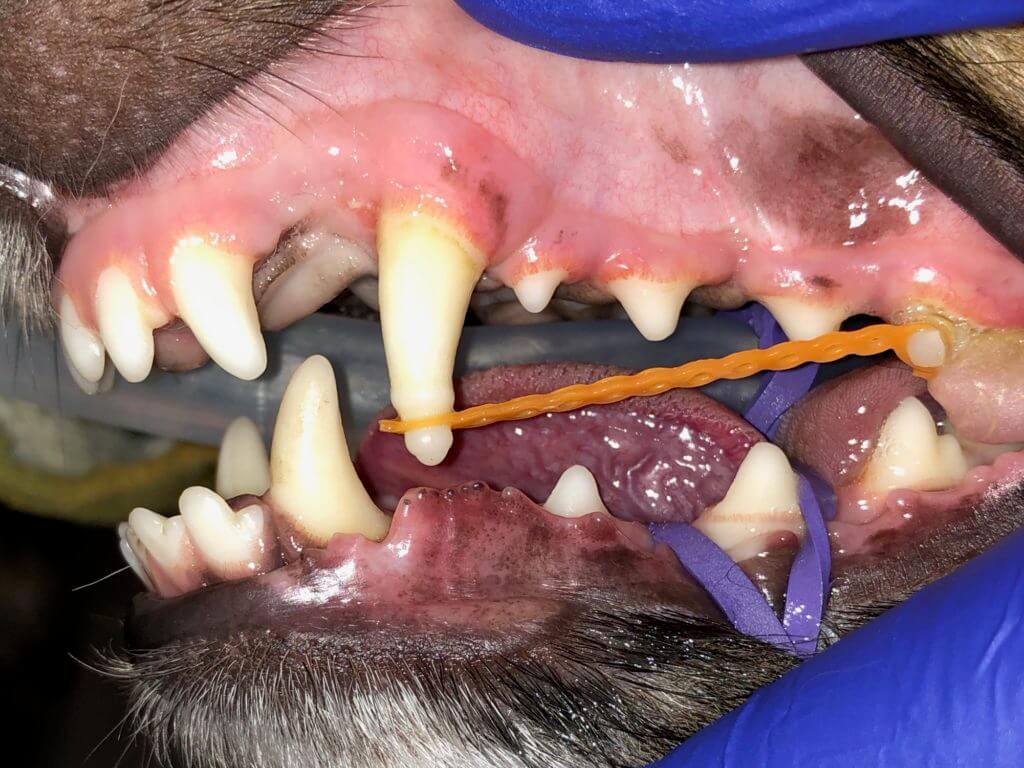
Yes, we can do braces in pets! Braces are done not for cosmetic reasons but to treat or prevent subsequent pain or infection. One of the most common conditions to use braces are lanced canines—cases in which the tooth is pointing forward instead of down. Correction with orthodontics takes 3 weeks to 3 months depending on the amount of movement and the pet age. Pets tolerate the brackets and elastics very well.
When poor positioning of a tooth causes crowding and subsequent periodontal disease, moving the tooth to correct the problem may be the answer. This procedure is best reserved for strategic teeth like canines. Adjustments to the elastics occur weekly and sometimes the brackets need to be relocated as in the initial and almost finished images below:
|  |  |
| --- | --- |
This is usually a genetic condition and breeding a pet after this procedure is unethical due to the risk of passing this condition on to offspring.
Orthodontic appliances can be used for other types of conditions as well. One example is an elastic training device to help keep the alignment of the jaw and support the muscles after the resection of part of the jaw following oral tumor removal.
- #### TMJ Problems 🐈
.jpg?width=288&height=192&name=closeup-portrait-of-orange-persian-cat-with-chubby-2022-11-14-06-05-27-utc%20(1).jpg)Pets that have problems opening and closing the mouth, or a clicking sound is heard may have temporal mandibular joint (TMJ) problems. Drooling or eating changes can be the only signs owners notice. Surprisingly many pets are good about eating even when they have pain. One or more types of imaging of the TMJ are needed. Depending on the findings, a surgical treatment or orthodontic elastics may be part of the therapy for recovery.
Temporal mandibular joint problems occur more frequently in pets than originally thought. Pets with only arthritis of the TM joint showed signs of issues less than half of the time, illustrating this is another hidden painful dental problem in pets. Feline patients with trauma are more likely to have TMJ involvement. 1.6 to 2 times more problems were found in dogs and cats with trauma when using CT to image the entire skull instead of two-dimensional radiographs.
In cases of trauma or difficulty opening/closing the mouth – it is necessary to have three-dimensional imaging to best evaluate the TMJ. 3D scans during all types of dental procedures may yield additional findings, decrease the duration of the procedure, and have significantly lower radiation.
For patients with TMJ problems, there are varied surgeries to treat the separate causes of the dysfunction. Not all TMJ problems require surgery to correct the problem either. Chronic pain from TMJ disorders is known to increase pain sensitivity to remote areas of the mouth. One must always be cautious of TMJ injury as months to years later the changes during the body’s attempt to heal the area may actually result in adverse effects.
For patients with oral tumors requiring resection of part or all of a mandible, the altered forces are more likely to cause TMJ issues of the functioning mandible (lower jaw). Elastic training of the muscles using orthodontics has been shown to help at least half of the patients as results are dependent on patient and owner participation with elastic training.
- #### Broken Teeth 🐕🦺 🐈
Chewing hard objects such as bones, antlers, hooves, etc... is the most common reason for tooth breakage. When the pulp is involved through crown or tooth root fracture, the only choices are extraction or root canal therapy (endodontics). While dogs have 42 teeth and cats have 30, only 8-10 of these teeth are needed for function. We wish to save these teeth through endodontics. This retains structure and function but the tooth is no longer living nor painful.
The center of the tooth is the blood and nerves, called the pulp.
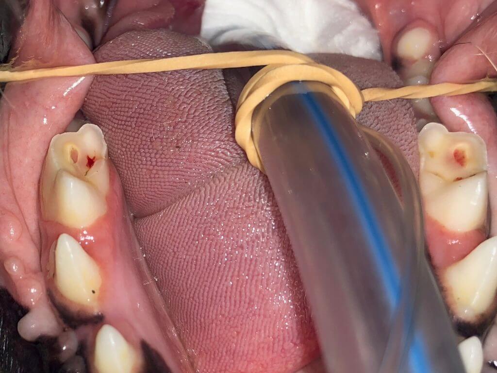
As stated above, once the pulp is compromised, a tooth must be extracted or have root canal therapy. Root canal therapy is known as endodontics. The pulp is removed and the bony walls of the tooth are cleaned with special tiny files. The area is flushed and disinfected, then dried. The root canal is then filled with gutta-percha (reminds me of rubber) to wall off this tooth from the rest of the body. The opening is then sealed with tooth-colored composite sort of like your dentist seals human cavities. While that all sounds simple enough, there are many techniques and expensive human supplies that are needed to complete the procedure well.
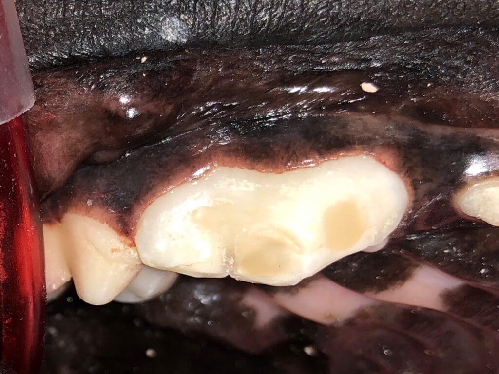
The bleeding broken tooth as shown above is usually not the case. In fact, the tooth didn’t even look like it affected the pulp:
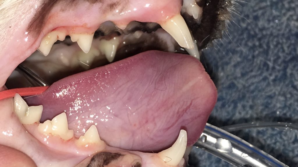
Sadly, a magnified anesthetic exam showed otherwise.
It is important to not ignore broken teeth as infection will cause problems locally and systemically.
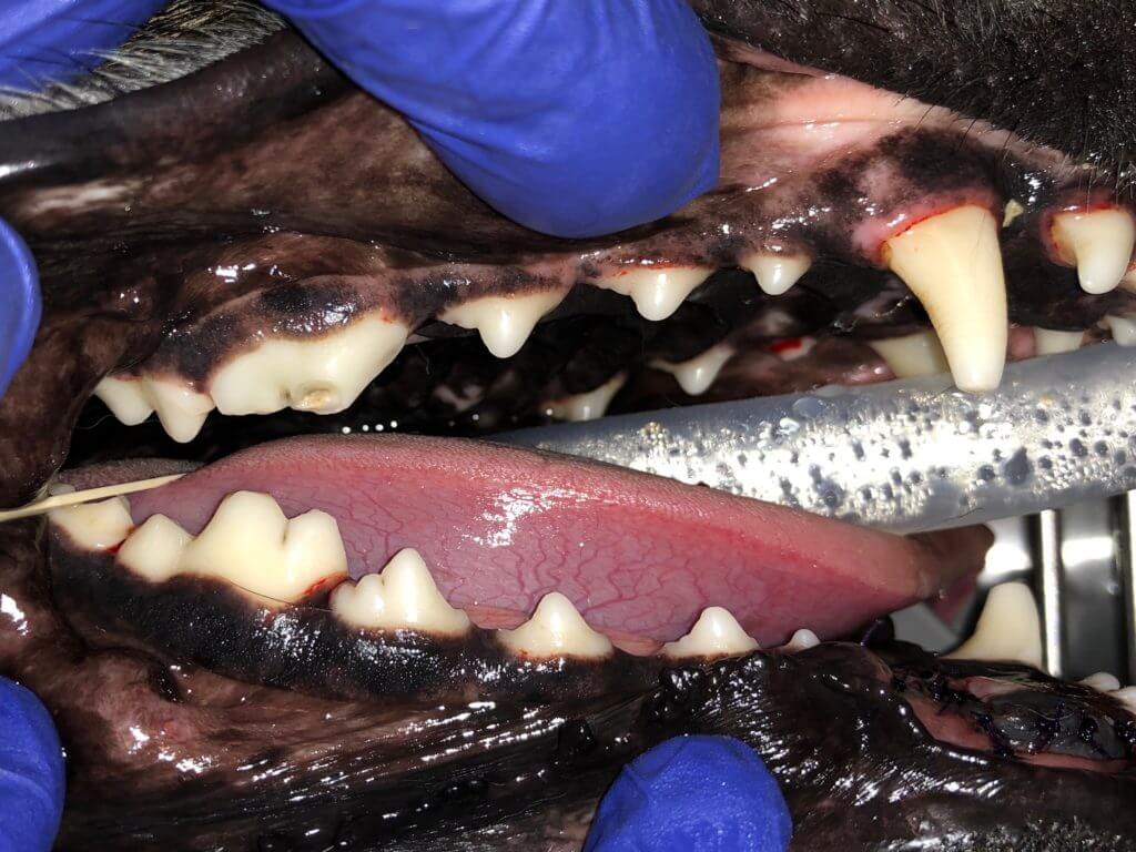
Each root of a tooth must be accessed and cleaned making some teeth like the three-rooted tooth below more of a challenge than others to treat. Holes need to be made into the tooth to reach each root. Although necessary, these holes weaken the tooth such that all teeth receiving endodontic therapy are best served with the application of a full metal (titanium alloy)crown. Once the root canal therapy has been completed, the pet is left with a non-painful, non-vital tooth that still functions. The good news is that endodontic therapy is around 95% successful!
|  |  |
| --- | --- |
- #### Open Root Tip 🐕🦺
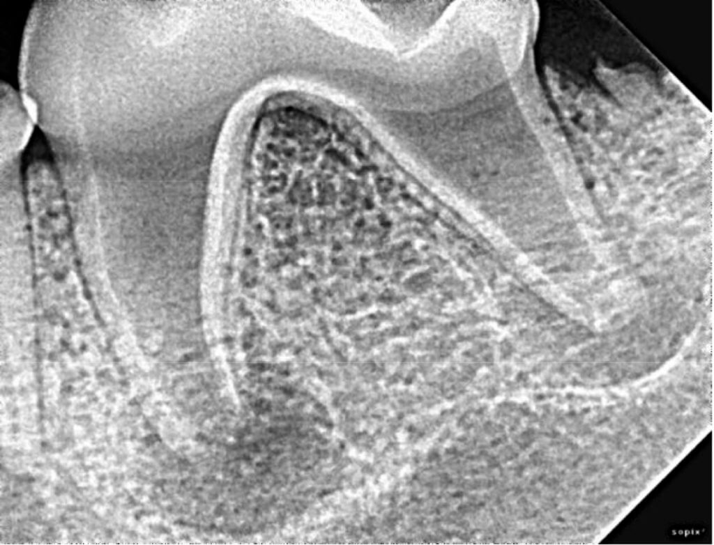
In some cases of trauma, the problem occurred while the tooth was still developing the tip of the root (apex). Other times disease of the tooth can dissolve the apex. These teeth still can be saved even with apical disease. They require a non-standard root canal therapy, best termed apexification that is performed over 2-4 separate anesthetic events to result in a structural and functional tooth without disease.
Non-standard root canal therapy is a situation in which the steps cannot be completed because the tip of the root (apex) is open preventing a tight seal, cannot be disinfected adequately, or cannot be filled properly.
This tooth was traumatized at a young age before the apex closed:
Disease at the apex of this [discolored](https://www.fampetvet.com/services/discolored-teeth/) tooth ‘dissolved’ the apex leaving it open:
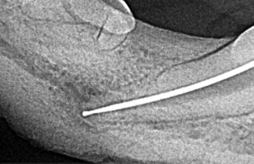
Special techniques and materials are used to close the apex and complete root canal therapy. This is known as apexification and results in a structural and functional tooth without the disease.
Although endodontic therapy is around 93% successful, in cases in which it is not successful, a surgical approach to endodontic therapy can be used. Other times simple bad luck with file breakage prevents appropriate filling of the chamber and may necessitate surgical access to complete an endodontic treatment:
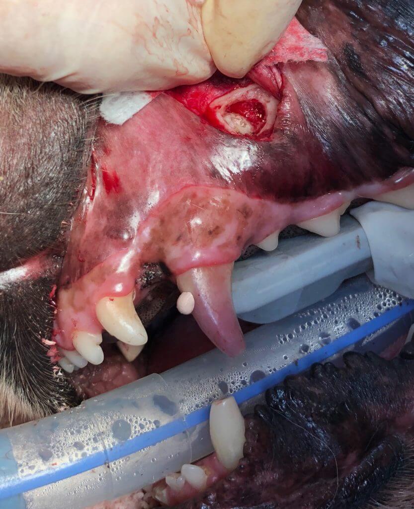
The good news is that there are many ways to save structural teeth!
- #### Recent tooth fracture 🐕🦺
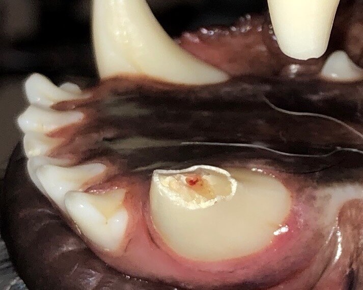
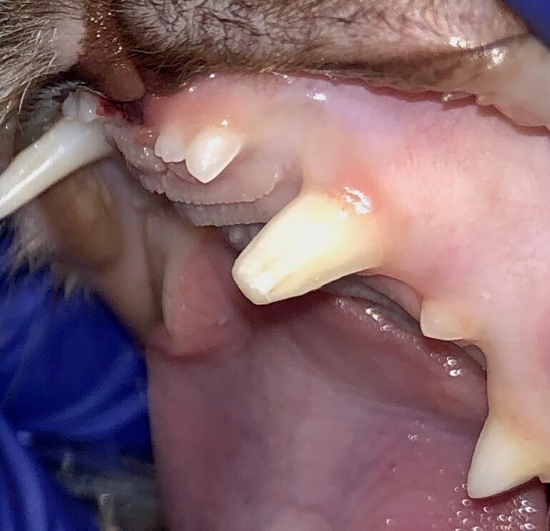
Sometimes a tooth will contact an object often during play and the time of tooth fracture will be known. If a tooth fracture is less than 48 hrs old on a dog older than 18 months, or less than 7 days old on a dog younger than 18 months, a ‘mini root canal therapy’ known as vital pulp therapy (VPT) may be performed. This preserves the vitality, structure, and function of the tooth and just makes it a bit shorter.
As stated in the image above, when conditions are right, we can keep the tooth living even when the pulp (blood and nerves) is exposed in contrast to a standard broken tooth and root canal therapy that results in a non-infected, non-vital, functional tooth.
This therapy may look and sound simple, but it is very technique sensitive and uses sterile instruments, and special biocompatible materials.
Case selection is most important to have equal success as standard root canal therapy. This ‘mini root canal therapy’ or vital pulp therapy (VPT) is limited to cases in which the pulp has been exposed for a short time. As time passes the likelihood of infection progressing deeper than the procedure removed pulp along with the body’s ability to respond in a positive manner results in less-than-ideal outcomes. If a patient was kicked in a cow pasture to fracture the tooth, the general time limits of less than 48 hrs for a dog over 18 months or less than 7 days for a dog younger than 18 months does not apply. In essence, it is important to know the age of the tooth and when/how the tooth break occurred.
In this dog, a hard object struck the tooth during play:
|  |  |  |
| --- | --- | --- |
In this cat, a fall luckily resulted in this tooth as the only trauma:
|  |  |  |
| --- | --- | --- |
Sometimes a tooth needs to be shortened due to a tooth contacting soft structures in the mouth usually for alignment reasons. Thus an intentional crown shortening can be the reason for this ‘mini’ root canal therapy. Vital pulp therapy is a shorter procedure than standard root canal procedures so it can be less expensive. In all types of endodontics, it is necessary to follow up with anesthetic imaging of the tooth in 6 months, then every 12 months thereafter.
Don’t despair if your pet’s broken tooth doesn’t fit the case selection for this procedure: as long as the gingiva is healthy, a fractured tooth can be saved by one of the types of root canal therapy: standard, non-standard, or recent fracture (VPT).
In this emergency case, call our [office](https://www.fampetvet.com/) or an [emergency](https://www.iowaveterinaryspecialties.com/) clinic telling them of the urgency of this case, the time at which the injury occurred, and the age of the pet. They will call my cell phone so I can meet you at an appropriate time, potentially after hours.
- #### Avulsed tooth 🐕🦺
There are cases of trauma in which the tooth is ‘knocked out’ or avulsed without breakage of the tooth root. These teeth need to be put into a glass of milk and can be reimplanted and splinted within 24 hrs. The blood supply to this tooth became disrupted so the pulp dies, but a follow-up root canal procedure one week later results in a structural and functional tooth without pain. In some cases, there is not enough bone nor can bone augmentation be sufficient due to surrounding disease, but in many cases, avulsed teeth can be saved.
When conditions are right, it is possible to save teeth that have been ‘knocked out.’ It is VERY important to put the tooth in a glass of milk and drive promptly to a veterinary dentistry center. The less time out, the better. In all cases, the injury must have occurred within 24 hours. There are exceedingly rare cases of teeth being repositioned within 30 minutes after the luxation and the tooth becoming vital again.
The trauma causing this situation often occurs in dogfights or road accidents (HitByCar). Sometimes the tooth is moved to the side (luxation/subluxation). Other times the tooth is pushed inward (intrusion). In all cases, there needs to be enough supporting bone and no or very little infection surrounding the tooth to allow the appropriate healing.
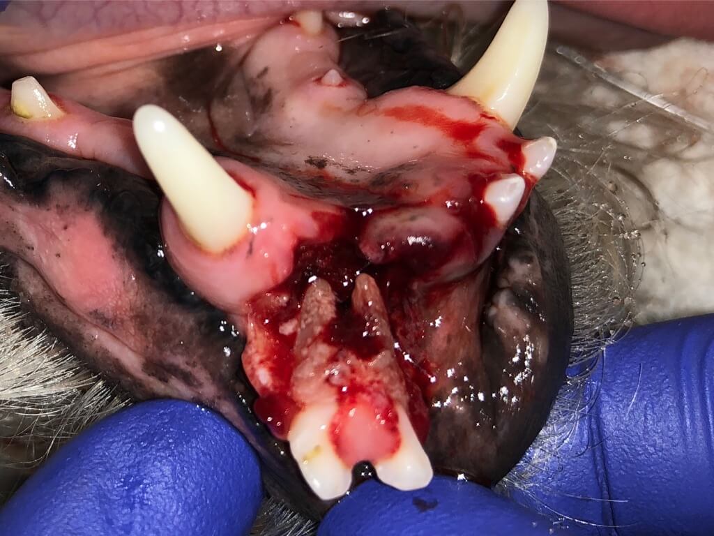
The tooth is splinted and a follow-up root canal therapy is performed one week later. Depending on each case, the splint may stay in place for up to four weeks, then be removed. In all types of endodontics, it is necessary to follow up with anesthetic imaging of the tooth in 6 months, then every 12 months thereafter.
In this emergency case, call our office or an emergency clinic telling them of the urgency of this case (keyword avulsed) and they will call my cell phone so I can meet you as promptly as possible.
- #### Discolored tooth 🐕🦺
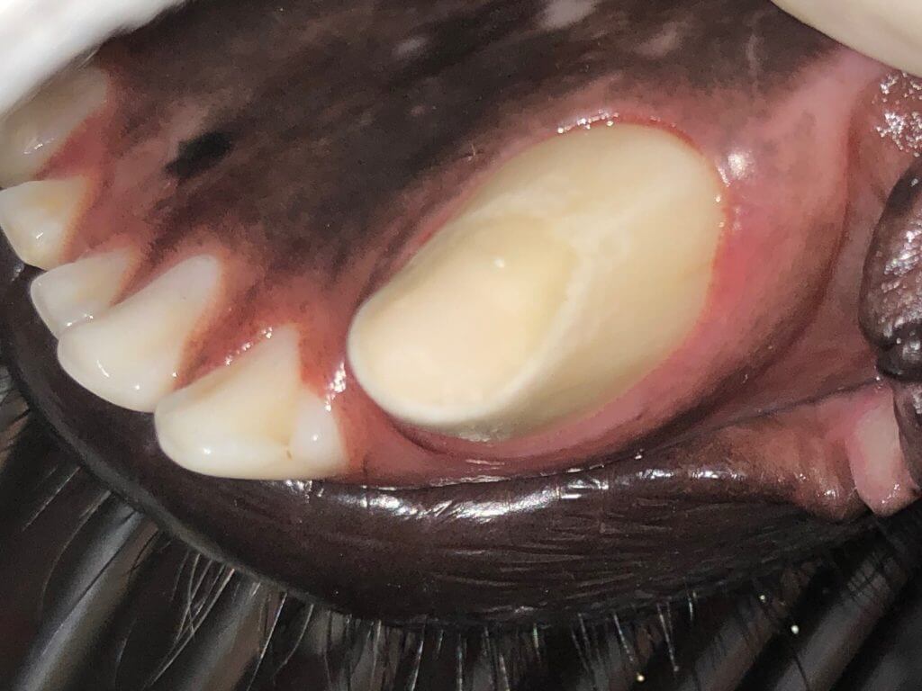
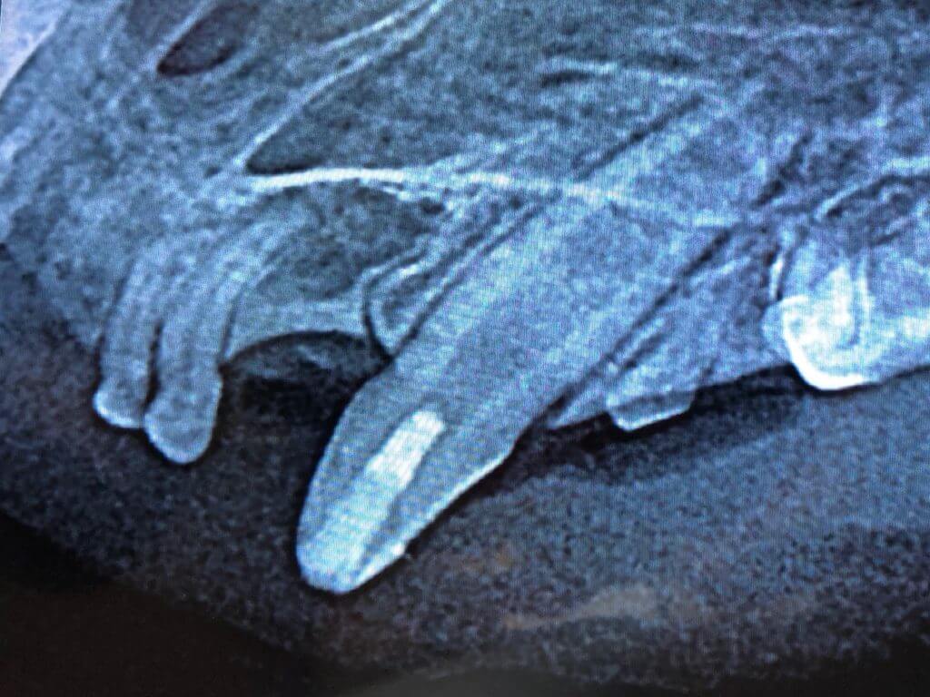
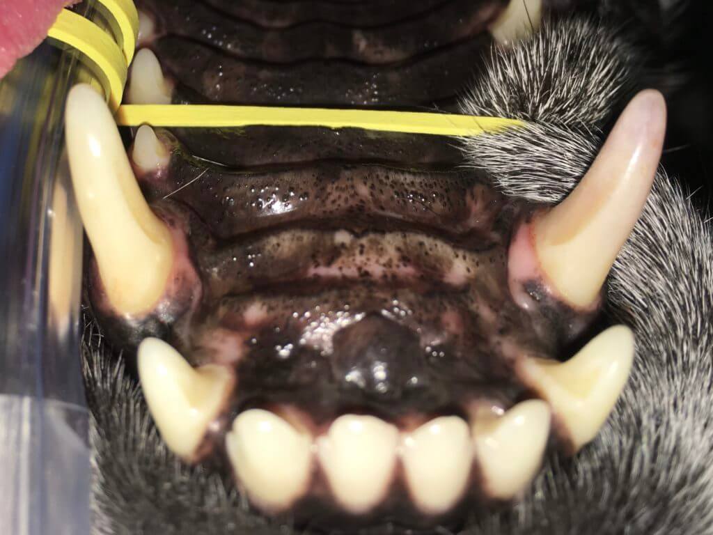
Almost all discolored teeth are dead. They are discolored because of the breakdown of the contents of the pulp leeches into the dentinal tubules discoloring the tooth. This is not just cosmetic as my father was taught, but a painful process. These breakdown products are a great place for bacteria to colonize exacerbating the problem. The only choices appropriate choices are root canal therapy or extraction.
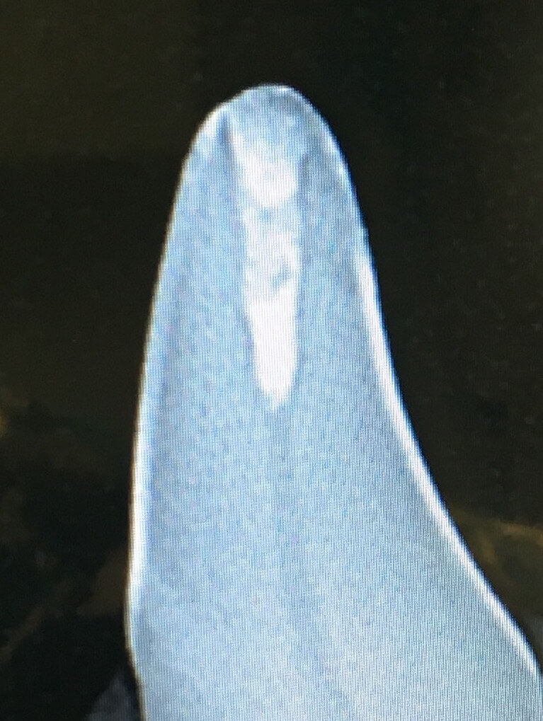
As seen in the image above and its accompanying x-ray, teeth age like inverse trees. Instead of laying down rings on the outer edge of the trunk, dentin is laid down inside the tooth adding structure and decreasing the pulp (tooth center nerve and blood vessels) diameter. When a tooth stops laying down dentin and similar teeth nearby do not, one sees a wider pulp chamber in the dead tooth. Other signs can be bone loss related to disease at the tip of the root, root resorption or narrowing of the pulp cavity due to calcification. One research paper states that only 42% of discolored teeth have x-ray signs, meaning that around 58% are still necrotic but don’t have x-ray signs yet.
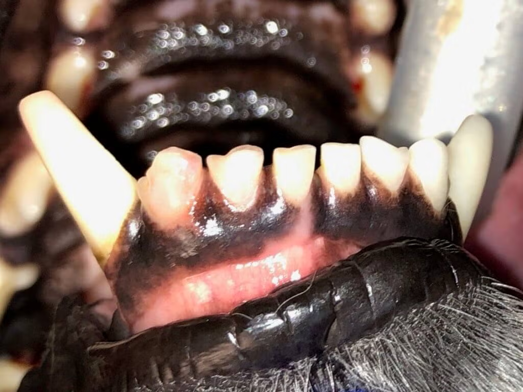
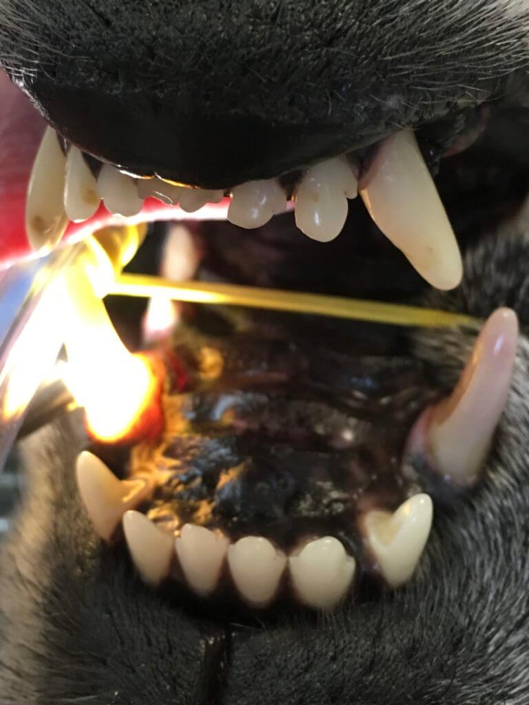
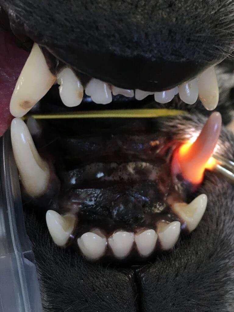
Another tool for evaluating a tooth to verify discoloration is called transillumination. It’s like candling an egg. A living tooth will have light shine through, while a dead one will not.
|  |  |
| --- | --- |
|  |  |
| --- | --- |
Although these images show obvious differences, it may not be this easy to tell. Transillumination alone cannot be used to make the judgment on treatment of a tooth. If one leaves behind a diseased tooth, the pet is left with pain or it will soon have pain at that site. There are [systemic inflammatory effects](https://tooth.vet/in-house-anesthesiologist?hsLang=en) known with diseases of the mouth, thus beyond pain, leaving behind disease is not wise. Extracting every affected tooth is preferable to leaving problems, but the most common teeth to become discolored are the canines and carnassials (the large important chewing teeth towards the back). There are a total of 8 of these structural teeth out of 42 total possible teeth in a dog’s mouth. The best course of action for the canines and carnassials is root canal therapy to save the structure and function but remove the pain and disease. One needs to be aware when deciding to extract these strategic teeth that they are losing almost all chewing function on that side of the mouth in the case of carnassials or removing a lot of jaw structure in the case of the canines. The most difficult tooth to extract in the entire mouth is a lower canine. This is one of the reasons that a common site for iatrogenic jaw fractures is adjacent to a lower canine extraction site. Root canal therapy must be considered, as it is best for the pet.
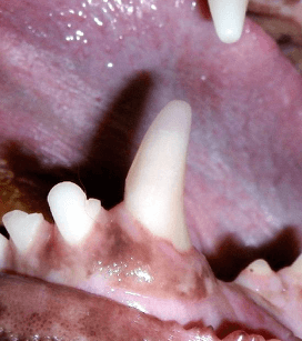The exception to this discoloration discussion is reversible pulpitis. When a concussive force occurs to the body, usually a bruise is formed and resolves it. When a concussive force occurs to a tooth, if a pet were lucky enough to have a ‘bruise’ and still have the tooth continue to live, the breakdown products would still leech into the tubules discoloring the tooth. The veterinarian can prescribe antibiotics (amoxicillin-clavulanate or clindamycin) and non-steroidal anti-inflammatory drugs (provided the pet’s bloodwork is appropriate) for 7 days. If the tooth’s color doesn’t return to normal after two or three months, you can assume it’s irreversible discoloration (pulpitis) leaving the only appropriate choices of root canal therapy or extraction. If the tooth improves, it must be followed with intraoral x-rays in 4-6 months, then every 6-12 months. The continued discoloration is likely an irreversible process and 42% of the time x-rays will verify it. It does take 4-6 months for x-rays to show mineralization change, thus answers will not usually be known right away.
- #### Full metal crowns 🐕🦺
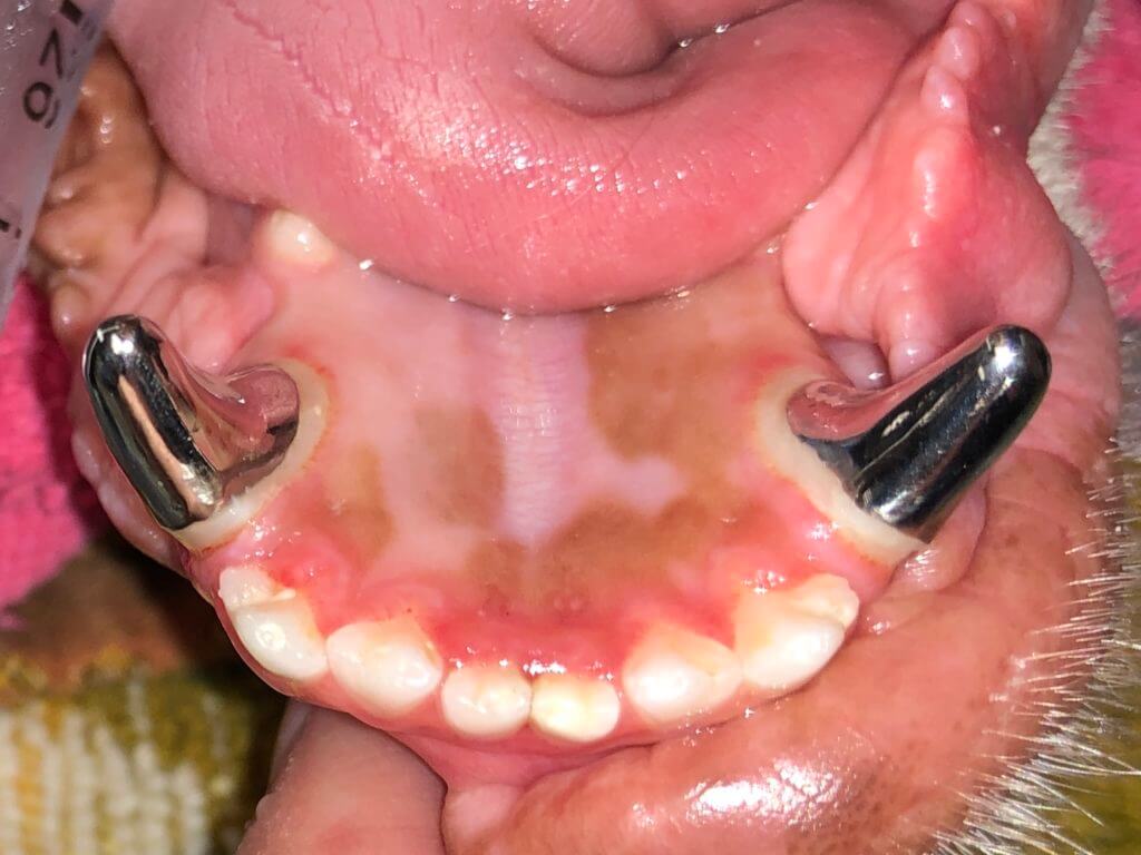
Any time a tooth is damaged, the structure is lost. If there is reason to believe the impact will keep recurring on this tooth (such as chewing hard objects), a crown should be considered. Some reasons for full metal titanium alloy crowns:
- Root canal therapy access sites can weaken the tooth.
- An uncomplicated crown fracture may become complicated with further chewing.
- Teeth with root canal therapy can be more likely to fail without crowns.
- These crowns return the tooth to full strength or stronger than the original.
Research shows that fractured and/or endodontically (having had root canal therapy) treated teeth are inherently more likely to break. One paper showed the treated teeth without crowns were up to six times more likely to break as compared to endodontically treated teeth with full metal crowns. The upper 4th premolar (shown above) is the tooth that is the weakest without a crown. Although full metal crowns are not required after a tooth receives root canal therapy, many choose to protect their investment by returning the tooth to its natural original strength or stronger.
Due to the physics of chewing in dogs and the fact that we cannot simply tell them not to chew on hard objects, the crown must be made of strong materials. Our office uses titanium alloy crowns for patients. Some veterinary dentists are trying zirconium (very durable and closer to tooth color) crowns. Porcelain crowns do not seem to last in most dogs.
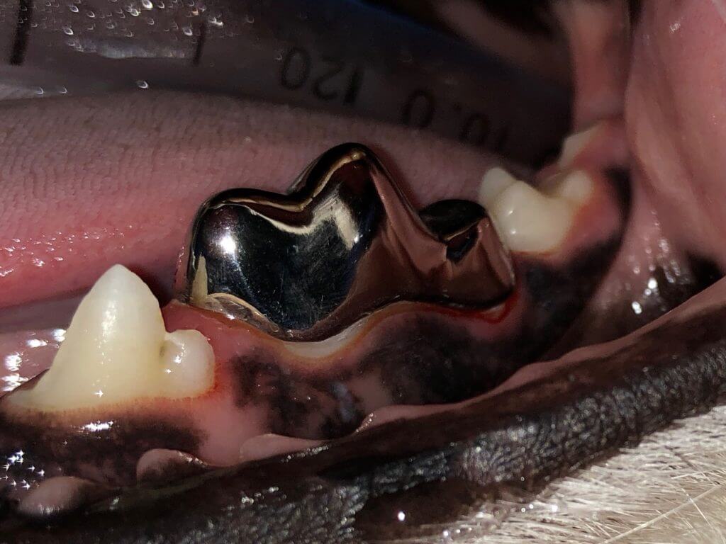
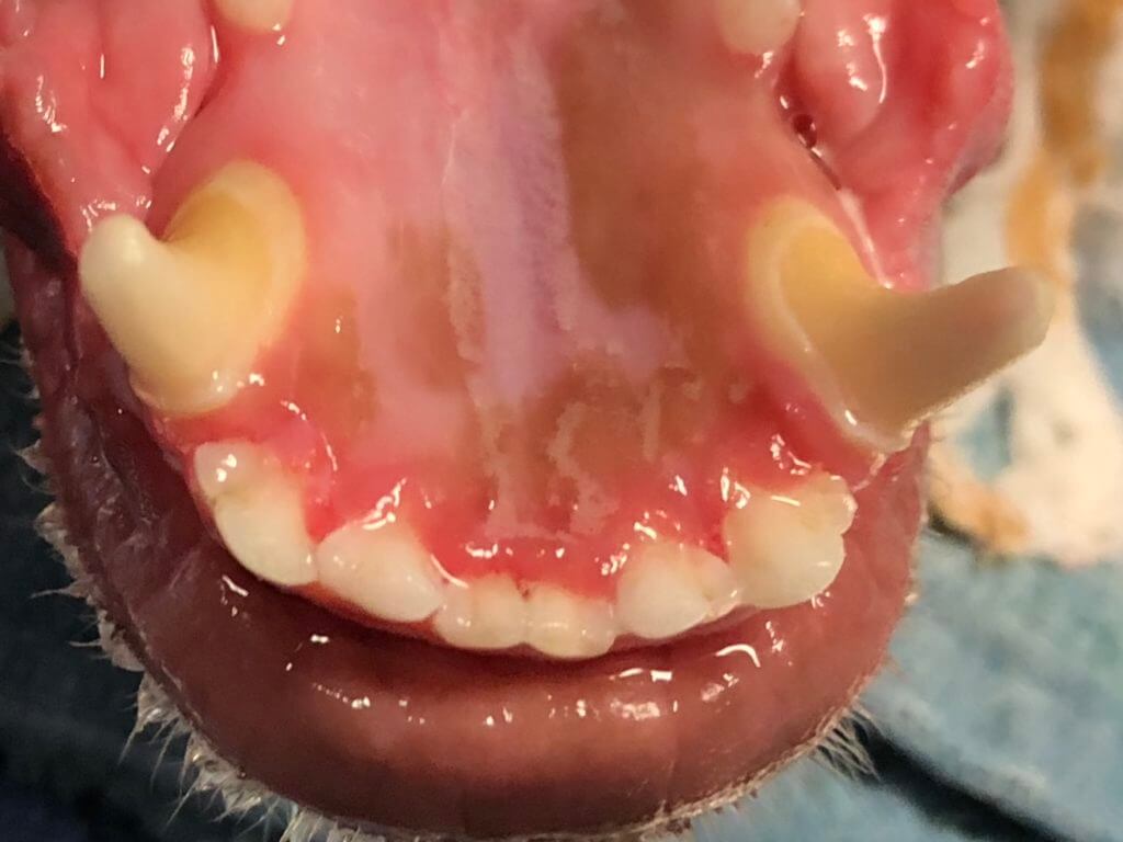
To allow the crown to fit, typically 0.7-1.2 mm of tooth is taken away with varying sizes and grits of diamond burs. The enamel of dogs is 0.1-0.6 mm thick meaning that only dogs that have sufficient dentin (usually by age 2) are good candidates for crowns. Additionally, as long as there is sufficient tooth height in proportion to its diameter, broken teeth or discolored teeth having received standard or non-standard root canal therapy are suitable for crowns.
Bones, antlers, hooves, and anything without flex can break teeth. The FDA even put out a warning to this effect. These are the primary causes of tooth breakage, root canal therapy, and subsequent full metal crowns. Appropriate chews and toys are any objects with flex. If your fingernail can indent the object, it’s okay. It’s even been reported that ice cubes break teeth. After root canal therapy, it is important to take away all items that can break this treated tooth as well as others. As many owners are surprised by what their pet finds to chew on, having a full metal crown gives strength and peace of mind should that occur. Continued chewing can still dislodge crowns so removing non-flexible chews/toys is still a good idea.
In some cases, chewing is happening on objects such as kennel bars and other items that cannot be taken away. Sometimes living/vital teeth are crowned to repair the damage and wear from kennel bars and/or uncomplicated crown fractures and give added strength for likely continued chewing.
- #### Crown fracture 🐕🦺 🐈
Sometimes a tooth will break and the pet will get lucky that the break did not expose the pulp (internal tissue of the tooth = blood and nerves). This results in exposed dentin. Dentin is porous (like lava rock). Saliva and bacteria infect the pulp through the dentinal tubules. If the radiographs (x-rays) show the tooth is not yet affected (~75% chance), a bonded sealant restoratove can be applied to seal the tubules stopping the problem. Six months later, follow-up radiographs are needed to verify early infection had not already started unseen and monitor success.
Tooth fractures are one of the most common dental problems seen in dogs. These fractures can be complicated(involving the pulp which is the center tooth nerves and blood vessels) or uncomplicated (break usually removing enamel, exposing dentin, but not pulp). Bones, antlers, hooves, and anything without flex can break teeth. The FDA even put out a warning to this effect. Chewing on bones breaks teeth. Appropriate chews and toys are any objects with flex. If your fingernail can indent the object, it’s okay. It’s even been reported that ice cubes break teeth. In some cases, chewing is happening on objects such as kennel bars and other items that cannot be taken away. A lucky pet will have the dentin exposed but not the pulp. Dentin exposure is painful. The odontoblastic processes (nerve endings) are exposed to fluid from the mouth. In addition to this pain, there is a risk of infection.
To seal the tubules and improve cosmetics, a restoration can be performed.
On x-ray this recent tooth fracture was fine for the moment:

So a restoration was place to seal the dentin, stopping pain and new infection.
Because the trauma that caused the uncomplicated crown fracture may actually cause irreversible pulpitis or bacteria may already be in the dentin but has not shown up as infection, intraoral x-rays under anesthesia are needed in 6 months, then every 12 months thereafter. As every pet needs at least annual dental preventative care, the every 12-month evaluation should not be a problem. X-ray changes can lag behind disease occurrence by 4-6 months, as there must be enough mineral change to see the problem on x-rays.
Occasionally the body can form reparative dentin. Waiting for this to happen is not a good idea, but should a darker area that is not soft when pressed with a sharp shepherd’s hook probe be present, treatment of the uncomplicated crown fracture is not needed. X-rays to get a ‘snapshot’ of tooth health under the gumline are still needed.
Sometimes living/vital teeth are crowned to repair the damage and wear from kennel bars and/or uncomplicated crown fractures and give added strength for likely continued chewing.
- #### Jaw fracture 🐕🦺 🐈
Numerous locations and angles of oral fractures can occur. In patients' long bones, veterinarians are taught to correct these with screws and plates. We should not treat the mouth as a long bone, as screws will enter the tooth roots devitalizing them and creating more problems that were originally there. All jaw fracture cases should see a veterinary dentist so
that the repair can be done through non-invasive methods, correcting the fracture and saving teeth.
As stated above, oral trauma should be corrected through non-invasive methods to save teeth whenever possible. Most training programs for boarded veterinary surgeons do not teach wiring and acrylic methods of correcting jaw fractures as seen below:
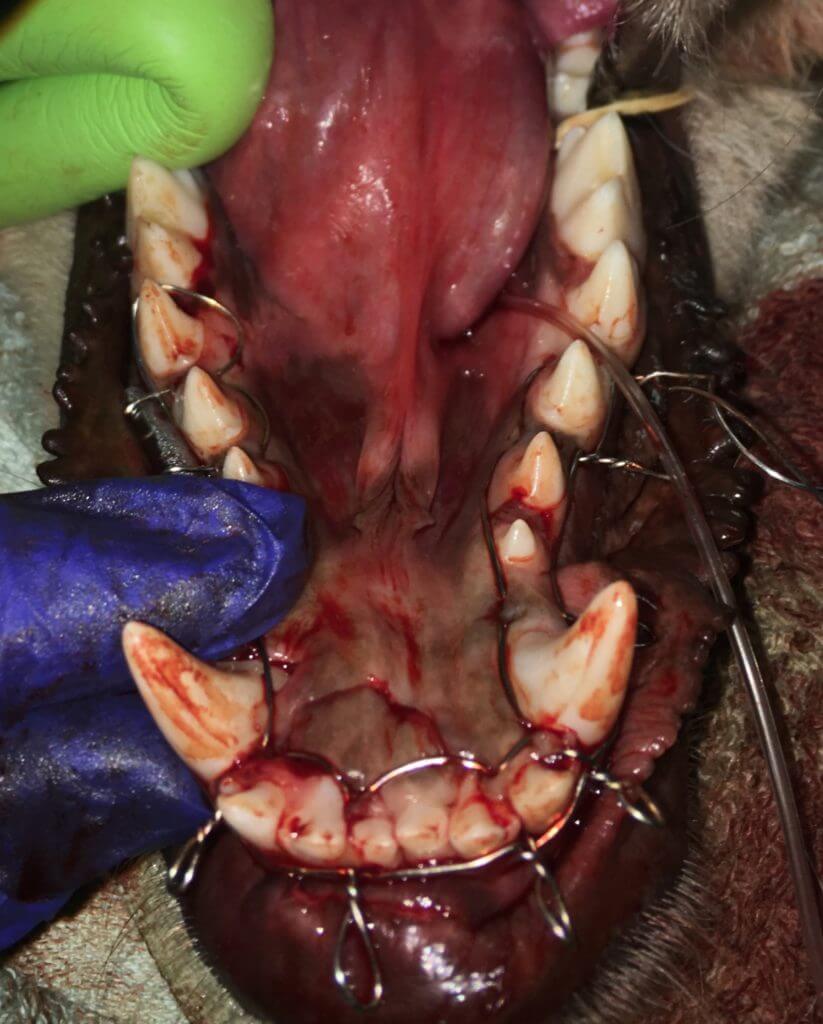
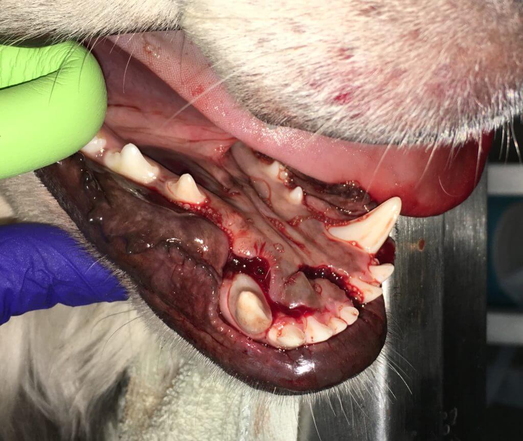
It is necessary to watch where the teeth contact and prevent the acrylic from contacting the gum tissues.
Wires may be used adjunctively to help in other areas of the mouth:
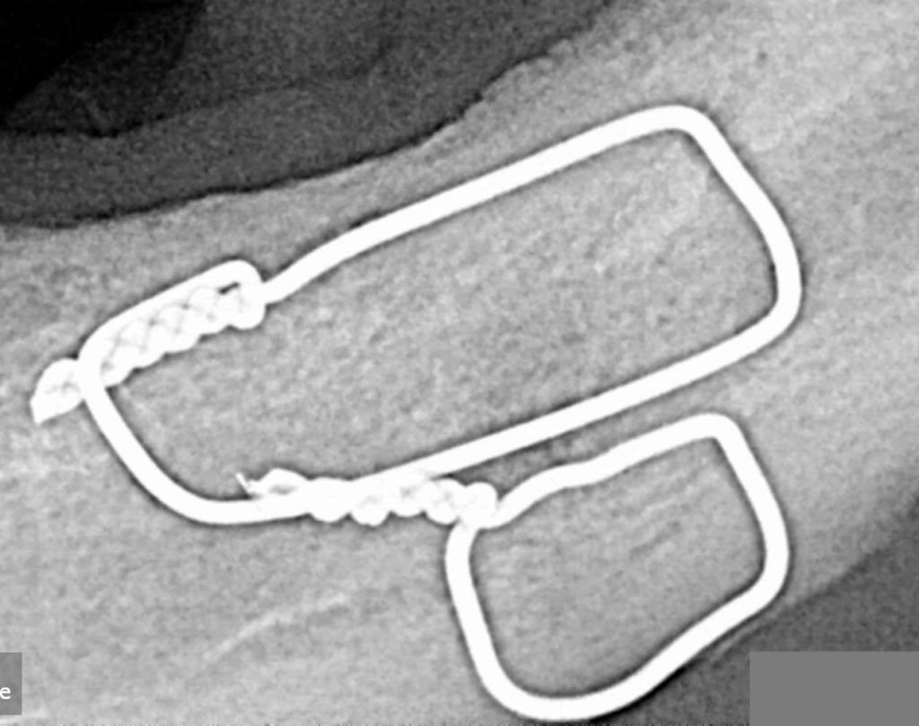
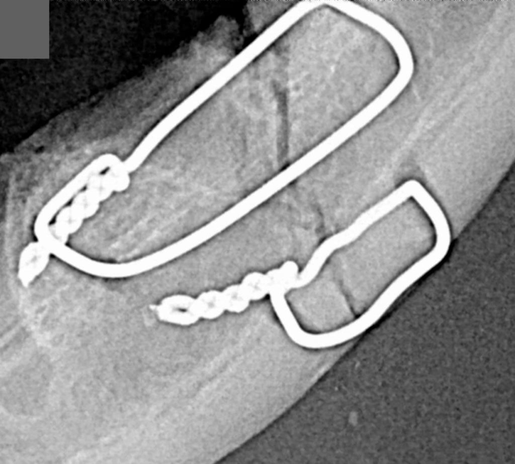
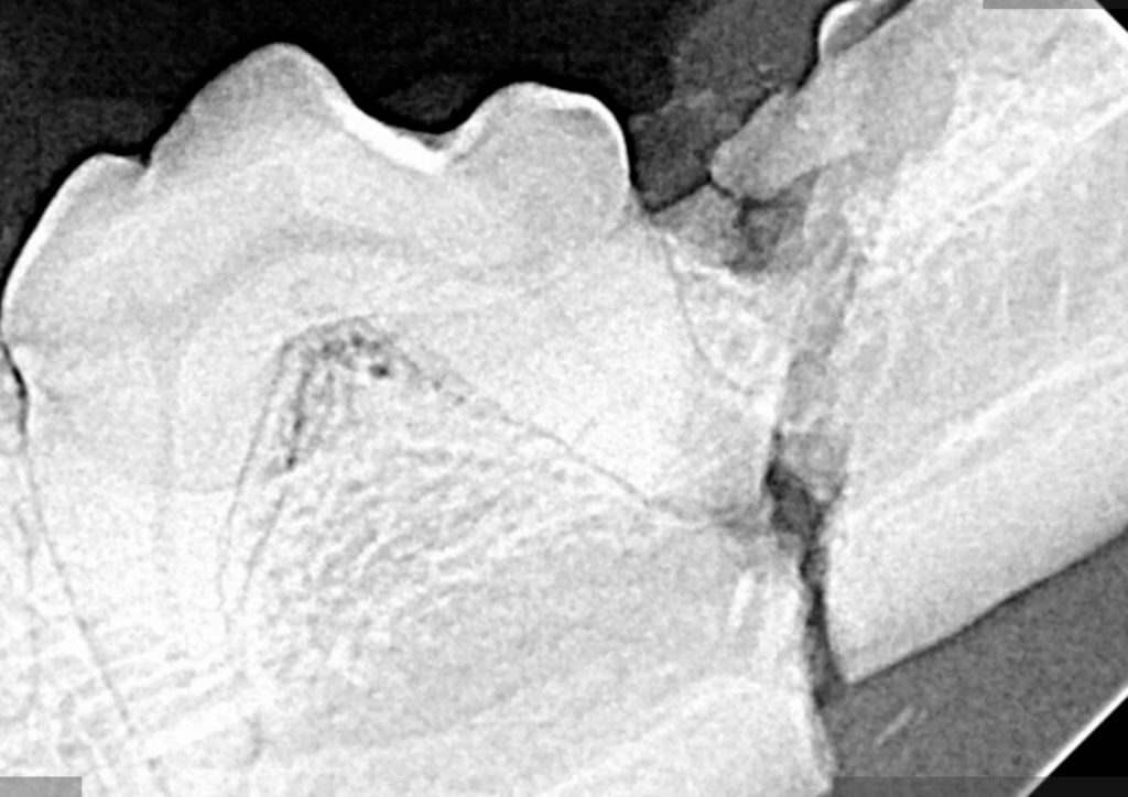
I urge extreme caution with fracture repairs in many general practices. Far too frequently, fracture repairs fail due to gum and/or bone infection that was present prior to fracture or occurred secondarily to improper technique.
Repairs of the jaw by a veterinary dentist allow for follow-up root canal therapy when indicated. Sometimes parts of teeth can be removed while others saved (hemisection). In all cases, keeping the structural teeth (canines and carnassials) when possible is best for the patient.
Cats are more likely to fracture the front and back parts of the jaw:
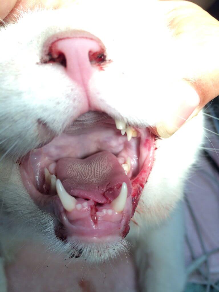
Studies have shown 1.6 to 2 times as likely to find additional injuries with 1mm or less CT slice images for cases of trauma. 3D imaging is needed for all fracture patients. TMJ imaging is performed as part of this scan. Just because a facility has CT imaging, it does not mean the detail is adequate for accurate diagnosis and treatment. Please ensure micro CT imaging is available. The imaging available in our facility is 3D cone beam computed tomography containing soft tissue as well as bone algorithms with true data instead of interpreted data. The detail is as thin as 0.09mm and uses less radiation. Most traditional CT units use about twice as much radiation and have typical slice thicknesses of 4-6mm = 20+ times less detailed.
Be cautious of symphyseal separation and normal laxity in a cat. This is one possible ‘fracture’ that may truly not be. Here is a video of normal wide symphyseal movement in a cat. Intraoral radiographs or 3D imaging need to be used to help check for the need for repair. How the mouth closes, patient history, and concurrent disease are also use to help guide treatment decisions.
- #### Cleft lip/palate 🐕🦺
During embryonic development, a defect can occur causing clefts/openings in the nose, lip, and/or palate. This allows food and fluid from the mouth to enter the nasal passages causing issues. Special surgical techniques are used to close the defects, as there is often insufficient tissue to close in a standard surgical fashion. These cases may sometimes require multiple surgeries to correct the problem but the outcome usually makes great improvement in the life of the pet.
Clefts of the lip and/or palate have been reported to occur in ~1 for every 1000 live beagle births. Due to the fact many puppies with clefts are euthanized early in life the true incidence across breeds is unknown.
Surgeries to repair clefts are varied but can be successful. Due to an average of 1.75 additional cm of bone missing on average under the visible soft tissue cleft defect, 1) repairs may require multiple surgeries and 2) 3D imaging is invaluable.
Studies have shown animals with clefts have at least one additional anomaly such as ear issues or nasal issues. 3D imaging is necessary to find the additional issue as a study without CT found only 13.5% (vs 100%) had additional abnormalities. Just because a facility has CT imaging, it does not mean the detail is adequate for accurate diagnosis and treatment. Please ensure micro CT imaging is available. The imaging available in our facility is 3D cone beam computed tomography containing soft tissue as well as bone algorithms with true data instead of interpreted data. The detail is as thin as 0.09 mm and uses less radiation. Most traditional CT units use about twice as much radiation and have typical slice thicknesses of 4-6 mm = 20+ times less detailed.
Surgery can interfere with growth if performed anytime prior to 16 weeks of age. Many cases may need to find alternate feeding methods such as orogastric tubes until they reach sufficient age.
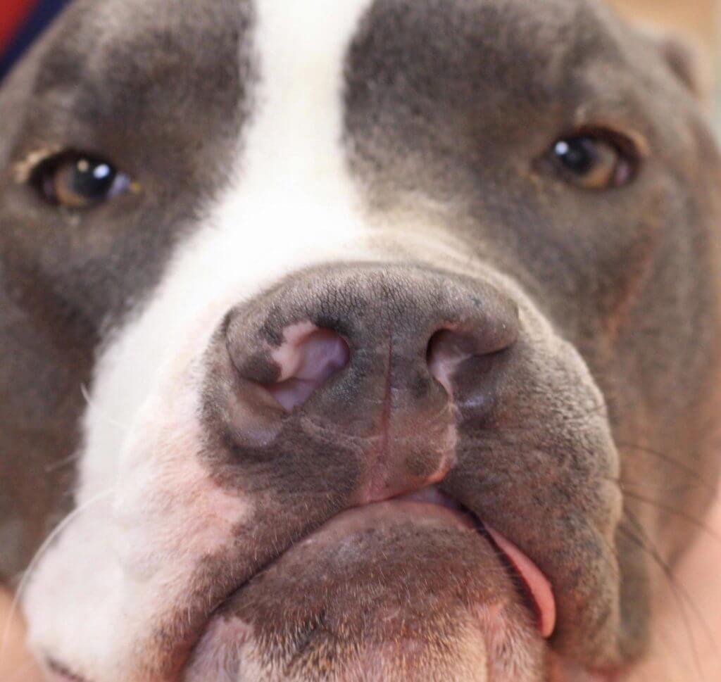
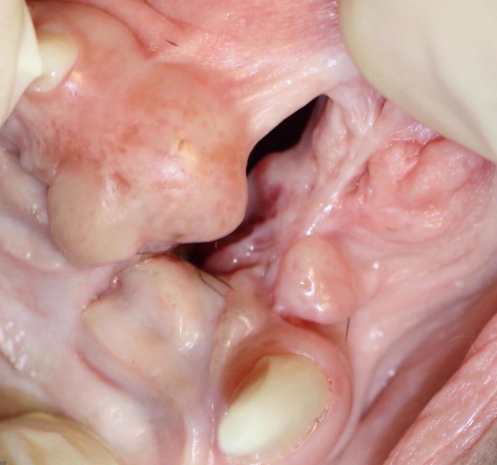
- #### Cavities in dogs 🐕🦺
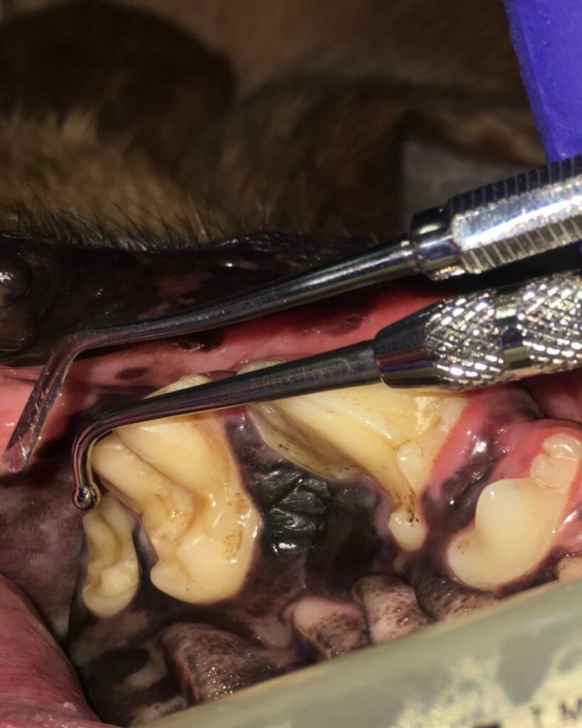
While not common, dogs can get true cavities just like, people! These are most likely to happen in the upper molar teeth that are shaped more similarly to human molars. Catching these soft pits early allows special restorative materials to be placed to correct the problem before it progresses into the pulp requiring root canal therapy or extraction.
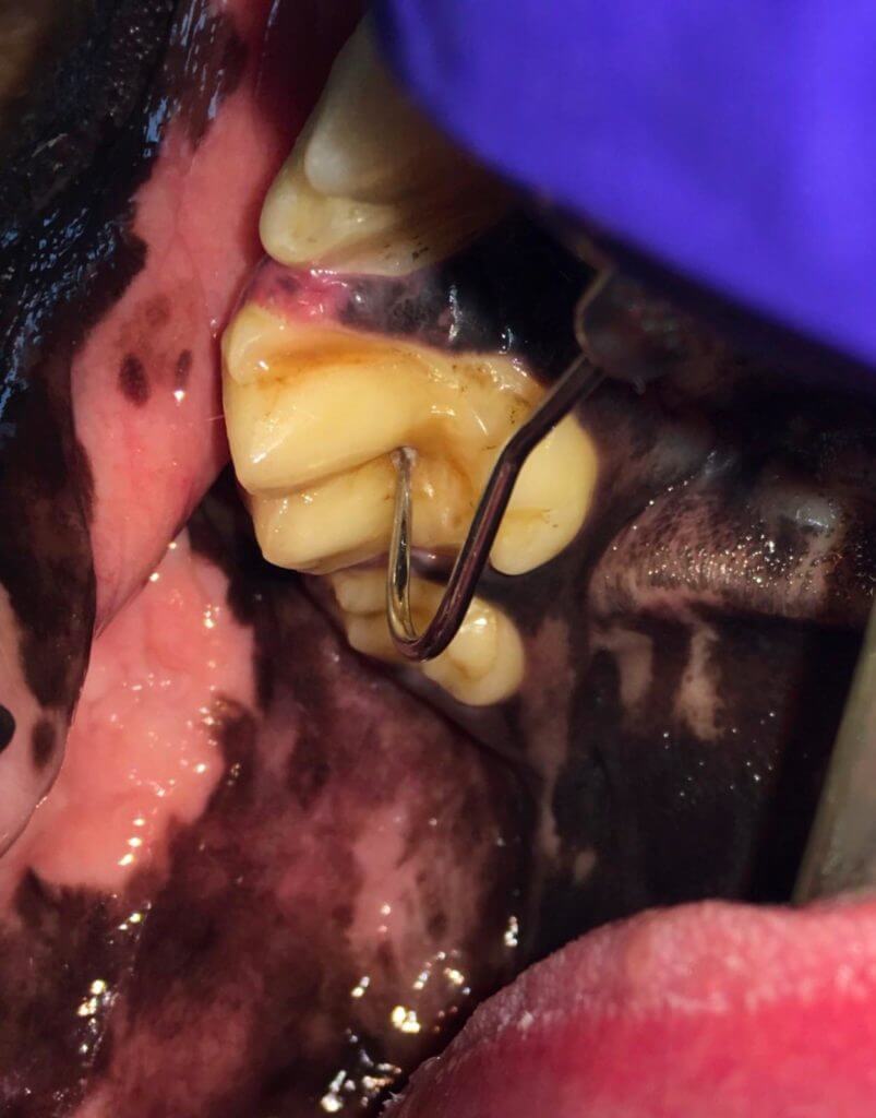
A soft indentation or ‘pit’ is the most likely finding. One of the first steps in the restoration process is to remove the diseased tooth material (dentin). Some dark soft spots are not these ‘cavity’ type lesions, but instead visually diseased pulp. Restorations alone are not appropriate therapy for endodontic (inside of tooth) disease. Should the disease or cleaning process progress to the pulp (innermost layer of the tooth), the tooth would require root canal therapy or extraction.
Check for soft spots:
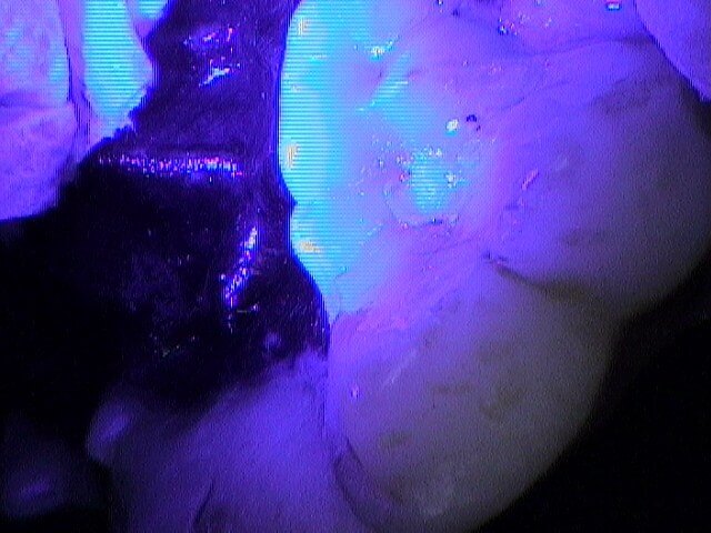
Special techniques are used to clean the defect. Certain materials are suited for the pressures of chewing, while others are not. Restorations require a curing light and smoothing instruments for each layer.
- #### Tooth resorption 🐕🦺 🐈
70% of cats get tooth resorption. These are often called kitty cavities but are unrelated to lack of brushing. Tooth resorption (TR) occurs as the body attacks its own teeth and ‘dissolves’ them. Because of the increased incidence of tooth resorption when inflammation is present when you treat TR through the extraction of teeth, you slow the recurrence.
Whole books could be written on tooth resorption (TR) alone.
Periodontal disease and tooth resorption are the two most common diseases in cats. Tooth resorption in cats has many names – kitty cavities, FORL, and more. The prevalence has been reported in different age groups ranging from 20-100%. One can say for certain that it is more prevalent in cats as they age (typically age 5+). One study showed 69% of mixed breed cats age 10+ had tooth resorption and 100% of pure bred cats age 10+ did as well. In the same study only ~ 20% of these patients had these findings on awake visual exams. Another study showed 2.4 times as many TR lesions were found on x-ray as compared to clinical exams.
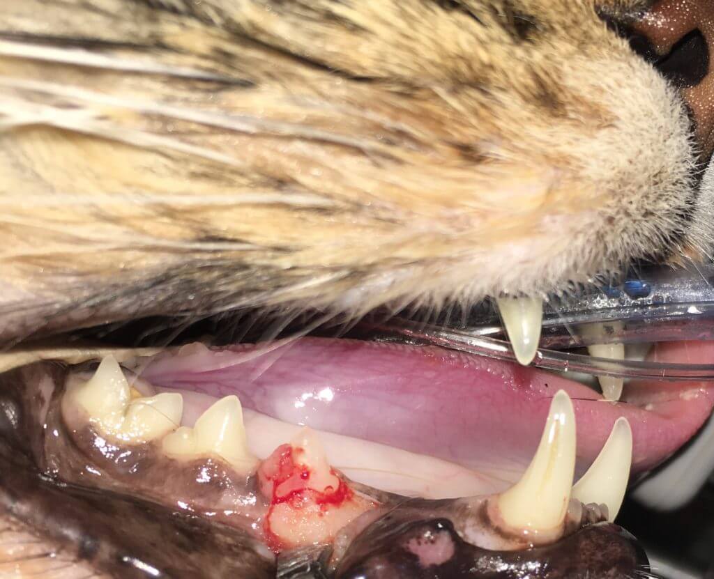
Here’s an awake finding that is certain to have a painful process:
The x-ray of this site under anesthesia:
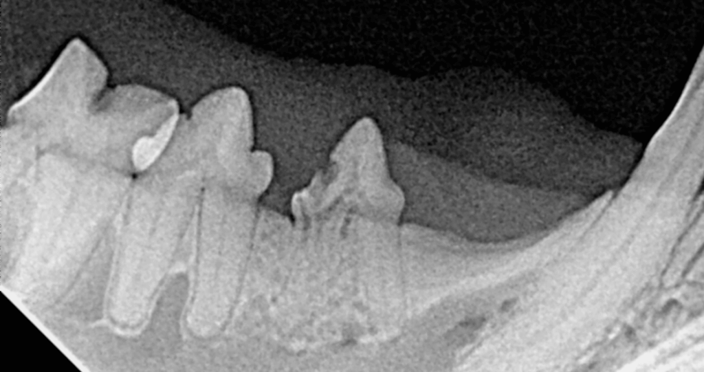
Revealing a focal ‘black’ spot of tooth (inflammatory tooth resorption) and loss of architecture of the root on the left (replacement resorption). A modified extraction technique (MET) can be used for the root on the left so that the remaining parts are below the gingival margin, but the root on the right must be removed in entirely or will continue to cause problems. Fragments of roots not having replacement resorption (aka retained tooth roots (RTR)) will not resorb. Tooth resorption is a painful often hidden disease usually happening along with periodontal disease. It is important to ensure your veterinarian uses intraoral radiographs or detailed 3D imaging for all cat dental procedures, as TR is not confined to the crown, but in fact, is a generalized recurring process. As stated above, due to the increased incidence of tooth resorption when inflammation is present, when you treat TR through extraction of teeth you slow the recurrence.
Some cats have canine teeth that are being extruded:
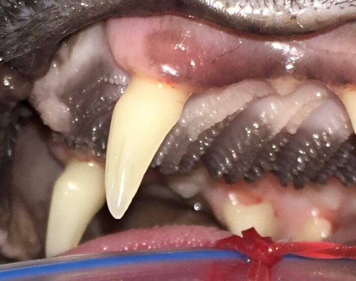
It is important to evaluate the whole mouth with intraoral x-rays in all of these cases as there is a significant correlation between extrusion of canine teeth in cats and tooth resorption. Other symptoms one may see on awake oral exam are buccal bone expansion (BBE) – this is enlargement of the bone usually surrounding the canine teeth in cats. (Green arrow)
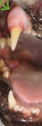
83% of BBE biopsy sites showed tooth resorption, another reason to ensure cats have the entire mouth evaluated with intraoral x-rays as well as probing for periodontal health measurements. Cats with inflammation in the back of the mouth are also likely to have tooth resorption as well.
(Caudal stomatitis):
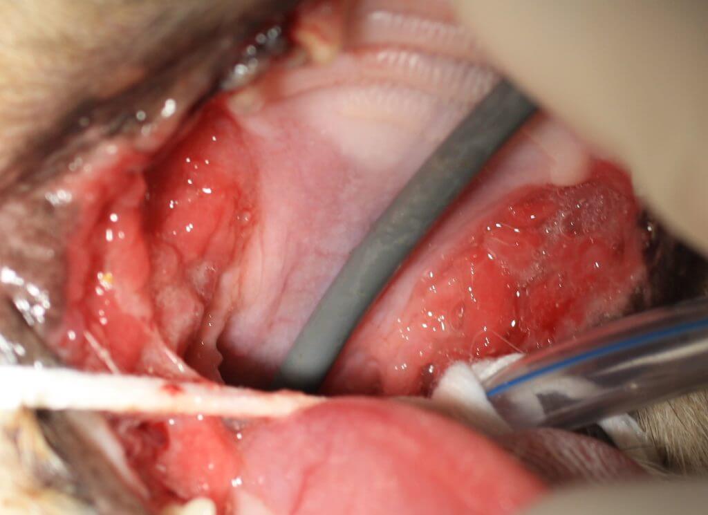
Feline Chronic Gingivostomatitis (FCGS aka stomatitis) is painful and medical therapy alone will not solve the problem. Check out the stomatitis section below to learn more.
In some cases, tooth resorption in cats and dogs are similar, but more likely they are different.
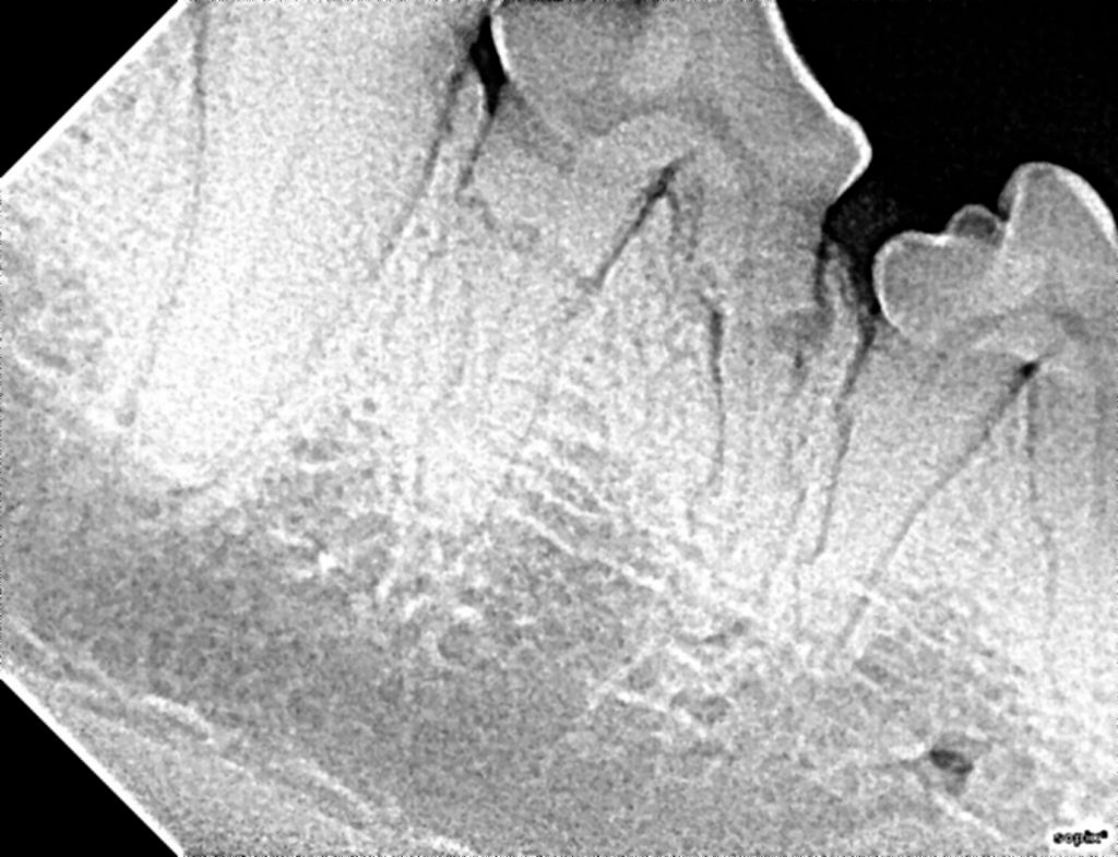
Dogs are more likely to get external root resorption:
This condition is the exception to the extract right away rule. This type of resorption tends to not be painful until it involves the pulp (but then it’s no longer external root resorption) or extends above the gumline. Should probing depths or tooth contour become abnormal, extraction then becomes needed.
Other less common findings are internal resorption:
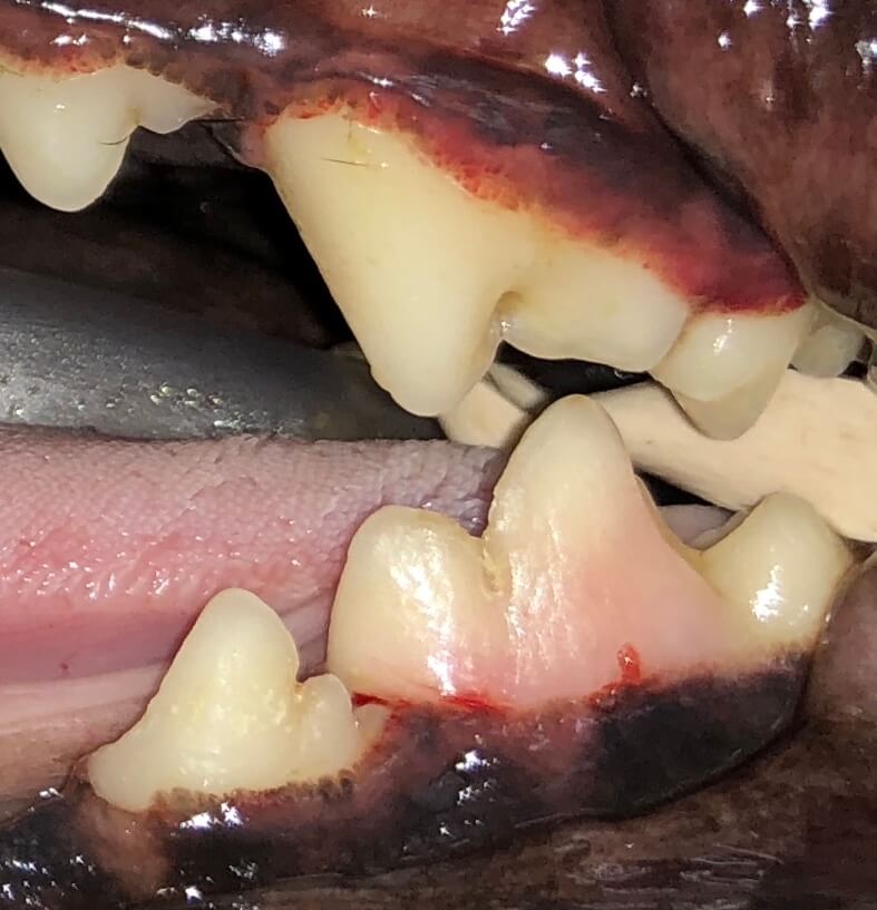
This problem is painful and requires extraction; we cannot watch and wait.
Tooth resorption can also occur uncommonly in exotic cheek teeth. Most of the time, the occlusal table needs to be equilibrated (aka floating teeth in horses), and the tooth will continue to erupt resolving the problem with frequent occlusal adjustments.
- #### Stomatitis 🐈
This term is used for widespread inflammation of the mucous lining of the structures of the mouth but should specifically be used only for cases that have caudal mouth inflammation/ulcerations. This is a painful condition that may have a relation to an underlying immune system issue. The gums often appear ulcerated and friable in addition to the caudal mouth.
While a study has shown all cases require extraction of either all teeth or all teeth behind the canines, when patients have been followed for longer than one year, those with retained canine still had recurrence of the condition. It's also important to note that if parts of the teeth remain (commonly retained tooth roots under the gum tissue), the condition may continue. It is very important to have detailed intraoral imaging to ensure the problem can be resolved appropriately for those that can respond.
As mentioned in the condition list above, tooth resorption is common in cats. Cats with stomatitis concurrently have tooth resorption and/or periodontal disease.
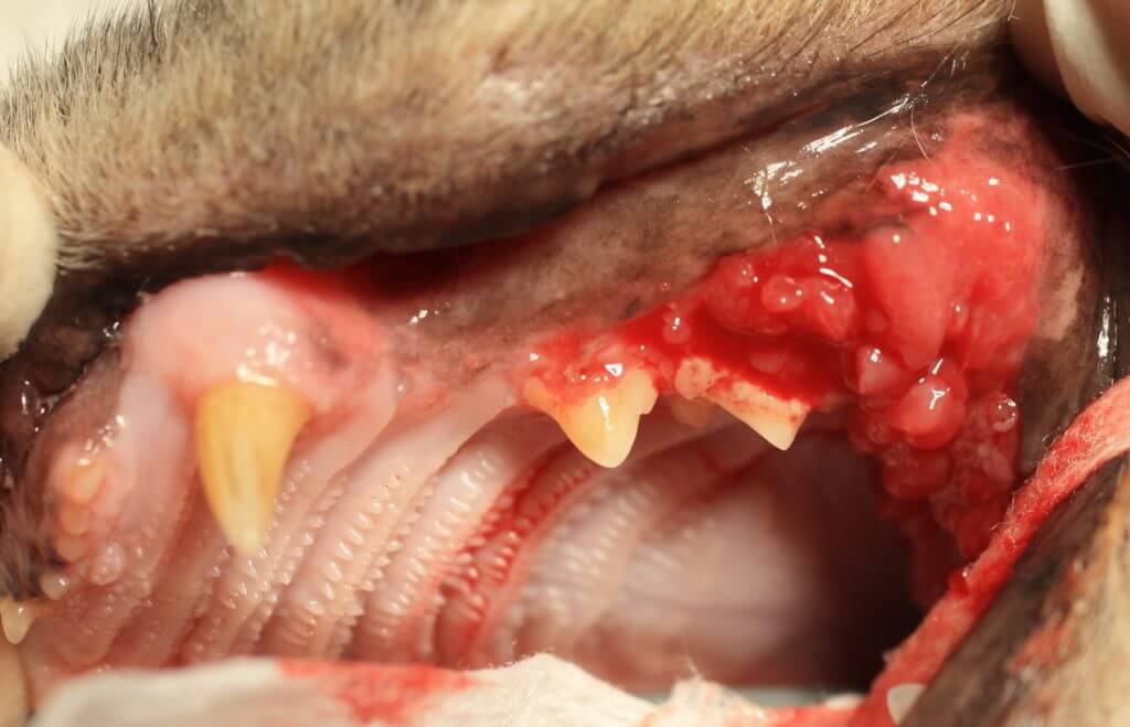
Should the inflamed ulcerated cheek teeth tissues NOT extend to the caudal aspect of the mouth (caudal mouth inflammation shown below), extractions in periodontally compromised and tooth resorption teeth should be performed, yet healthy teeth can remain. Once caudal mouth inflammation is seen, extraction of all teeth in the mouth is indicated even if they appear healthy.
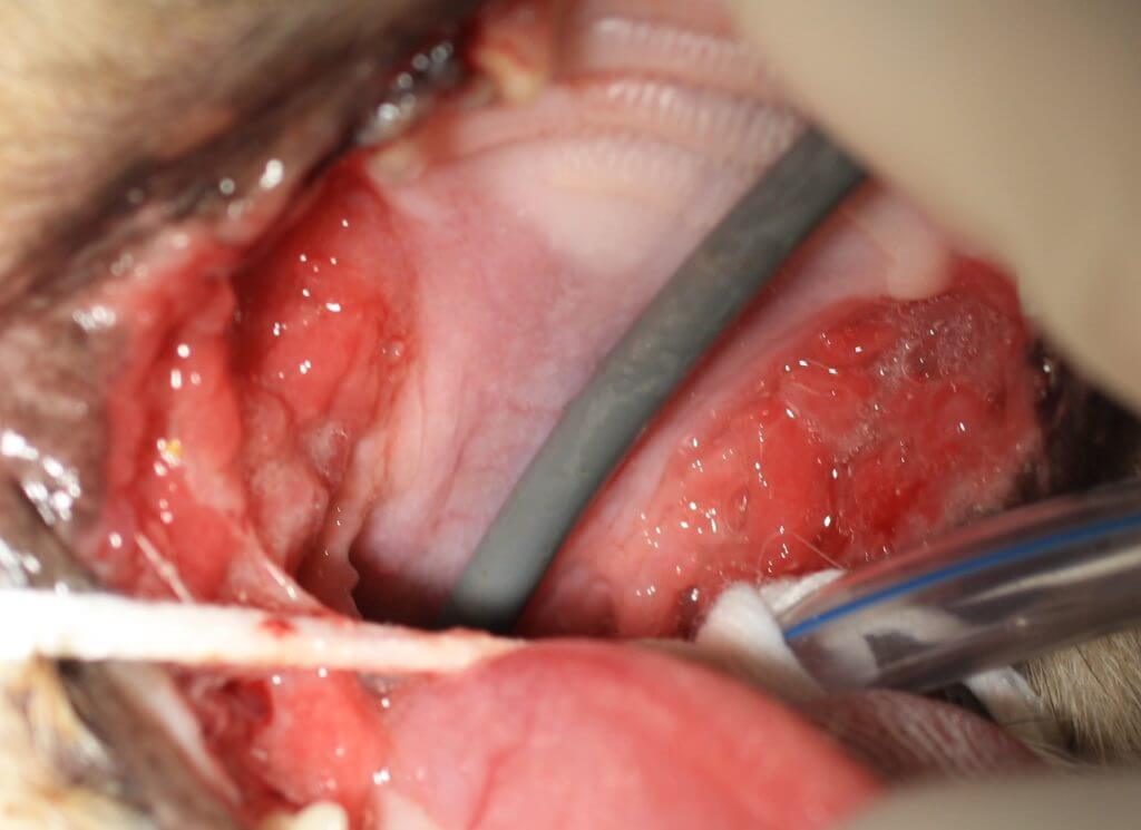
Some cases of caudal mouth inflammation are mild:
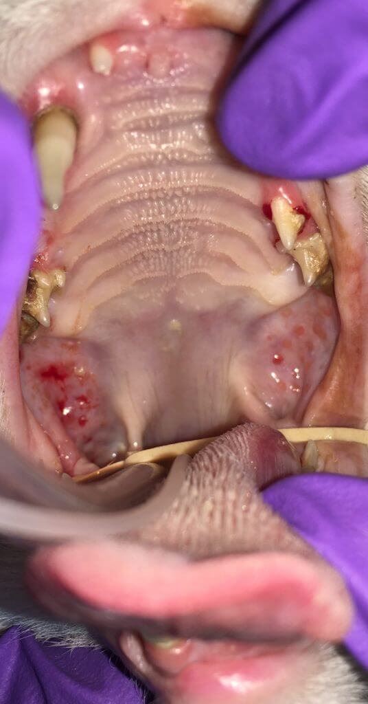
All cases of caudal mouth inflammation require full mouth extractions (FME) including all retained roots. Some believe that if the canines and incisors are unaffected, they may be retained. This is known as caudal mouth extractions (CME) as all teeth behind the canines are removed. While some have improved in the short term, all patients our office has seen with CME have had recurrence of issues. Another consideration is that some patients with stomatitis do not resolve. Others have advocated for medical management. A 2020 paper verified that all stomatitis cases should have medical management. The myth is that medical management will resolve the condition. Medical management is needed, yet is it important for medical management to occur along with surgery (FME). Medical management alone does not work. Sadly the ones that have had longer presence of symptoms +/- medical management without FME are less likely to fully respond once FME is performed. The sooner we get to removing the teeth (the plaque retentive surfaces, the more likely the cat is to have a desired outcome.
Many patients with full/caudal mouth extractions benefit from a few days of feeding through a tube as the mouth heals. While all of these extractions (FME) sound radical, they are necessary. Patients without extractions such as this do not resolve, as this is a condition with an underlying immune system issue that is thought to be an overreaction by the body to the plaque on the teeth.
Approximately 70% of cats with FME or CME respond well with either complete resolution (1/2) or significant improvement (1/2) still requiring adjunctive medical management. Sadly ~1/3 of stomatitis patients do not respond well to any therapy. An stated above, there may be a better outcome when full mouth extractions as first line treatment occur earlier in the course of disease.
- #### Extractions, special flaps 🐕🦺 🐈
Any time a tooth is damaged, we need to remove that painful tooth or preserve the tooth by another method (such as root canal therapy). There are specific teeth that should be saved if at all possible as these teeth provide structure and function to the mouth: all
4 canine teeth, the upper 4th premolars, and lower first molars (large chewing teeth aka carnassial). If teeth are damaged beyond saving, have tooth resorption, or are crowded creating disease, they should be extracted.
The above image explanation sums up the need for extractions well. The strategic structural teeth (the canines and carnassials) should be saved instead of extracted as long as the gum and bone surrounding the tooth margin (periodontium) are adequately healthy.
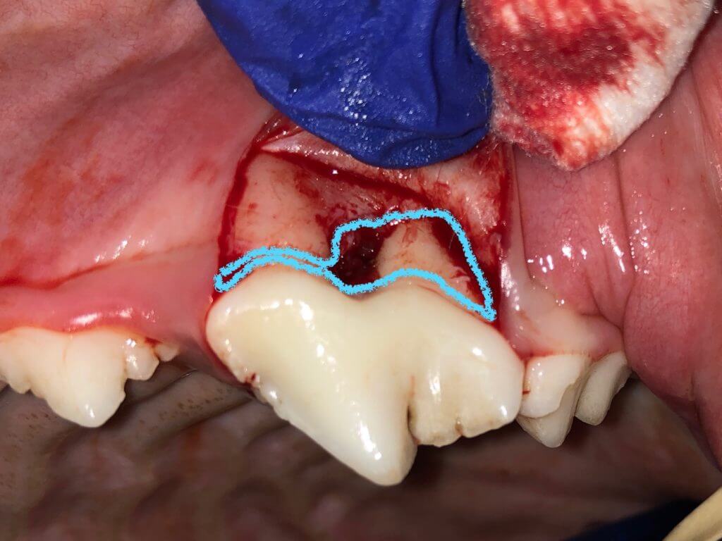
This tooth has a draining tract:
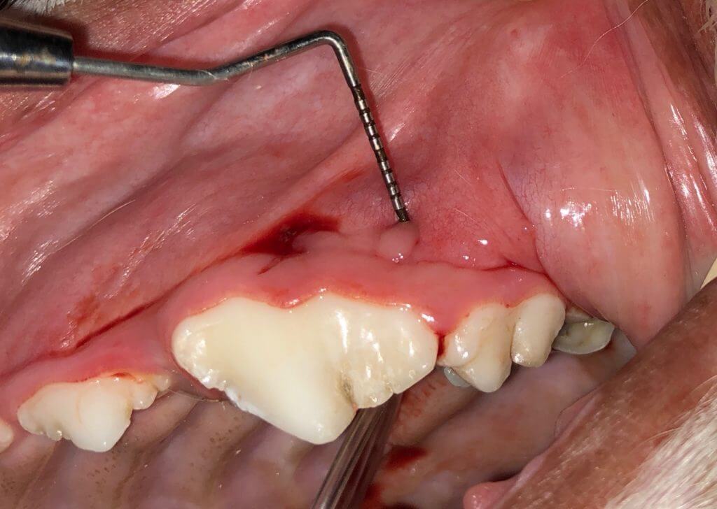
Much surrounding bone has been lost due to disease (outlined in blue). As the gum cannot reattach without supporting bone and the space between the two roots has missing bone, extraction is the best choice even though this is a strategic tooth:
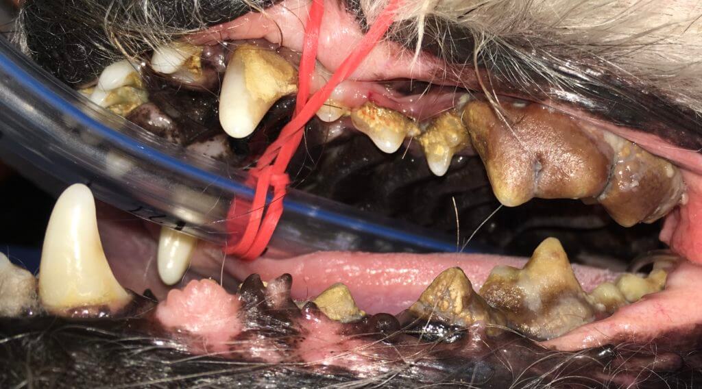
Dogs typically have 42 teeth and all but 8-10 have significant function. Teeth may be extracted as part of the therapy for a painful bite:
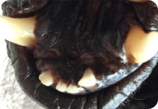
Tooth roots need to be removed in entirety (with the exception of replacement resorption) or may cause problems as shown below and in the baby teeth section.
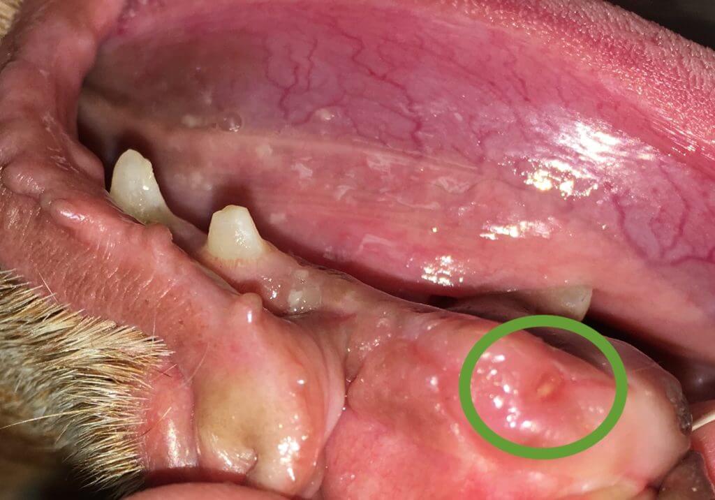
Extraction of teeth can still leave parts behind, so an x-ray after extraction is necessary to ensure the extraction is complete
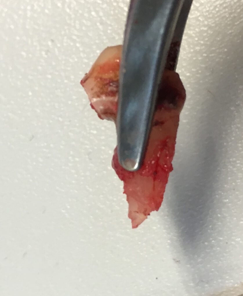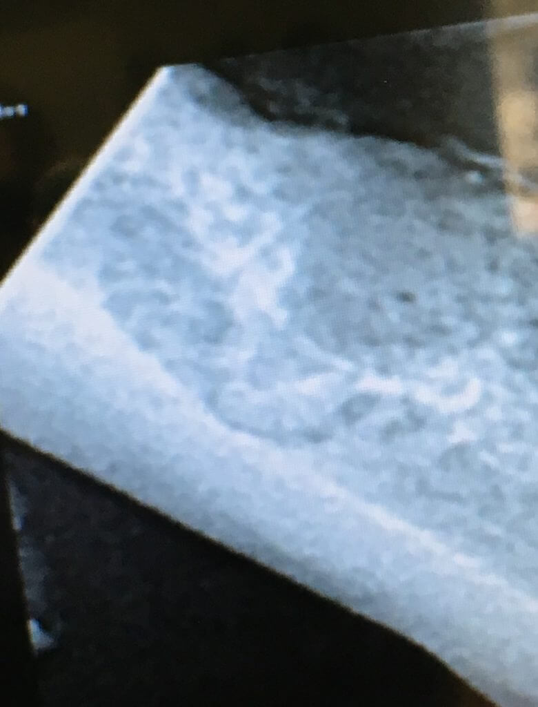
Sometimes we take out teeth that are not yet diseased but will cause it (rotation):
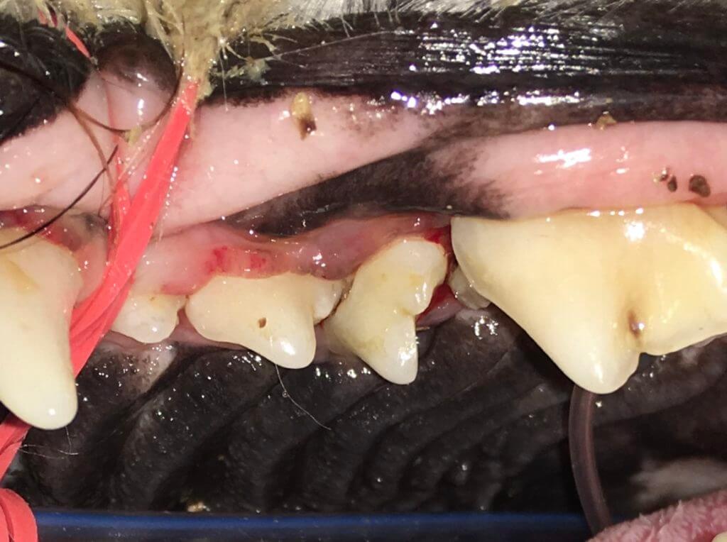
Other times teeth have malformations requiring extraction:
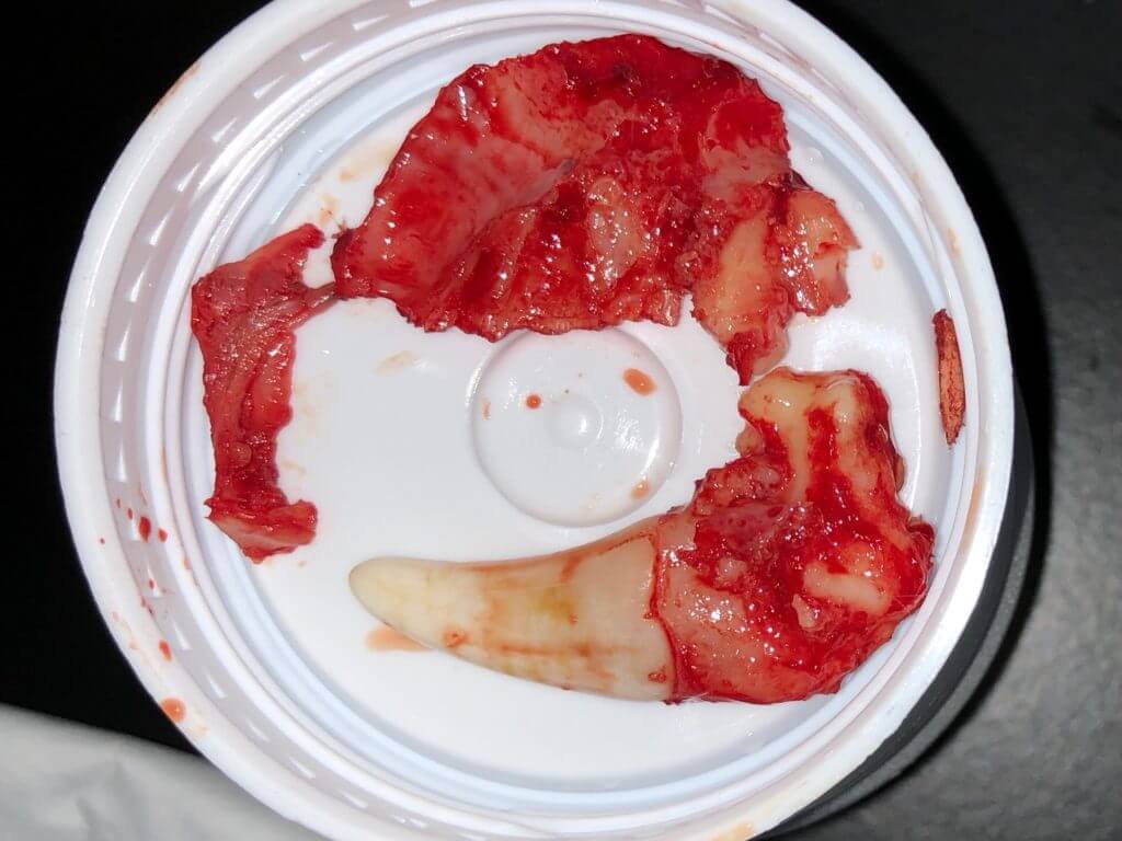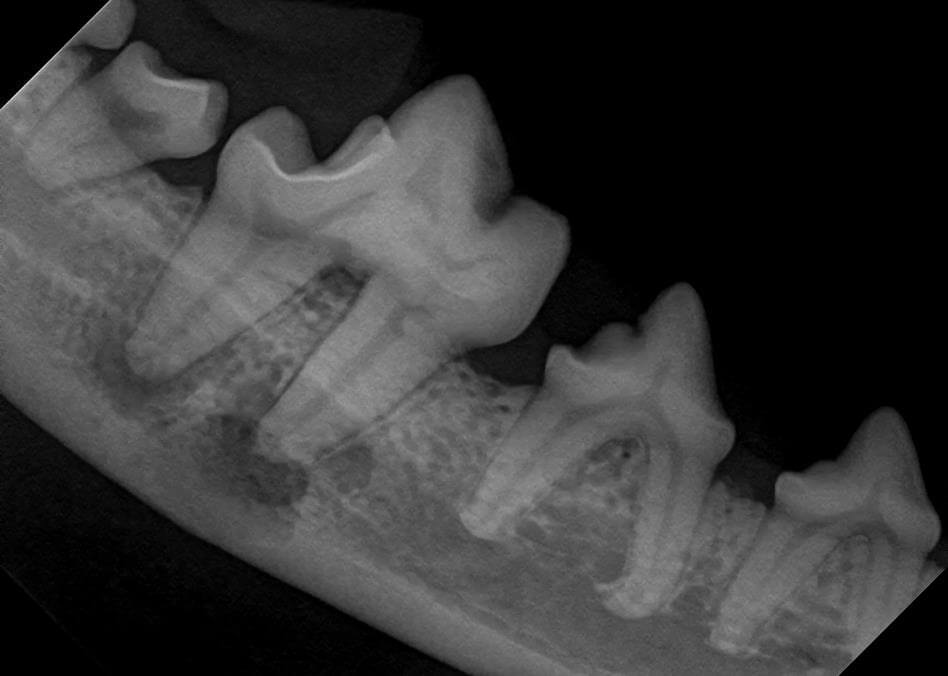
When extracting teeth, there is risk of jaw fracture when appropriate anatomy and forces are not observed or when the bone health is compromised as imaged above. While fractures can be repaired, some conditions require bone grafts or special techniques. The goal with these techniques is to save structure and in some cases, preserve teeth:
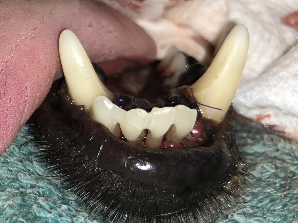
When performing extractions, oral surgical techniques must be used to close the site (unlike common practice in humans).
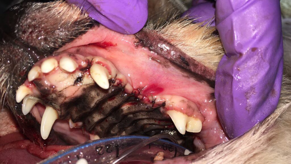
Won’t a pet have trouble eating with missing teeth? Dogs and cats do fine with missing teeth, but an opposing tooth does not have the natural cleaning action and may perpetuate periodontal disease. A missing opposing tooth can also result in trauma to soft tissues. Sometimes this can be corrected with tooth shortening, other times extraction is again required. Exotics with continuously growing teeth that require extraction, the opposing tooth often needs extraction as well.
As mentioned in the stomatitis post, this situation usually needs caudal mouth or full mouth extractions. These patients usually benefit from a few days of tube feeding through the esophagus as the mouth heals.
- #### Dentigerous cysts 🐕🦺
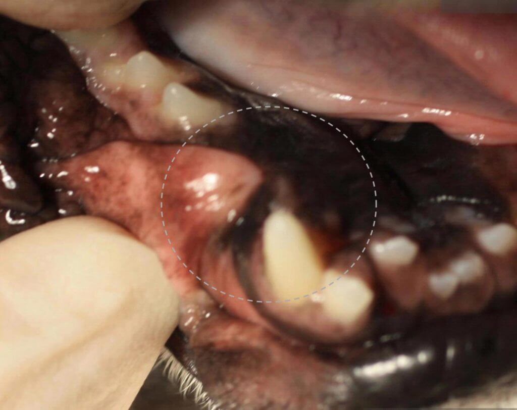
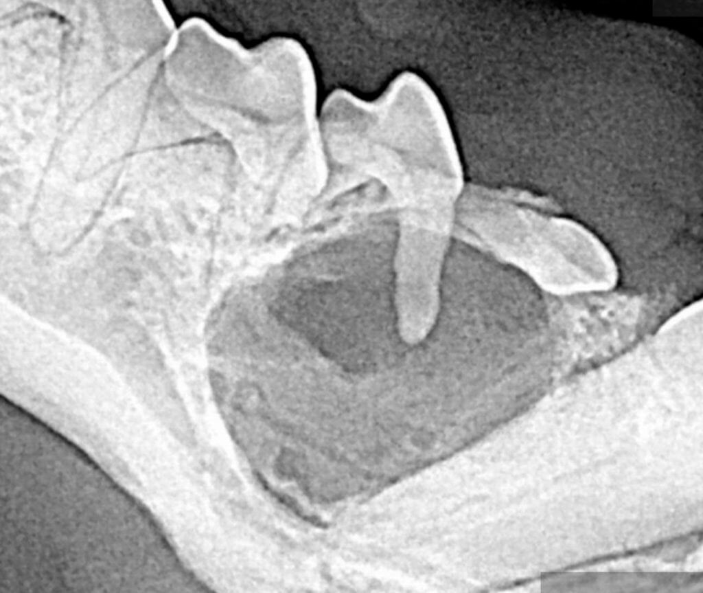
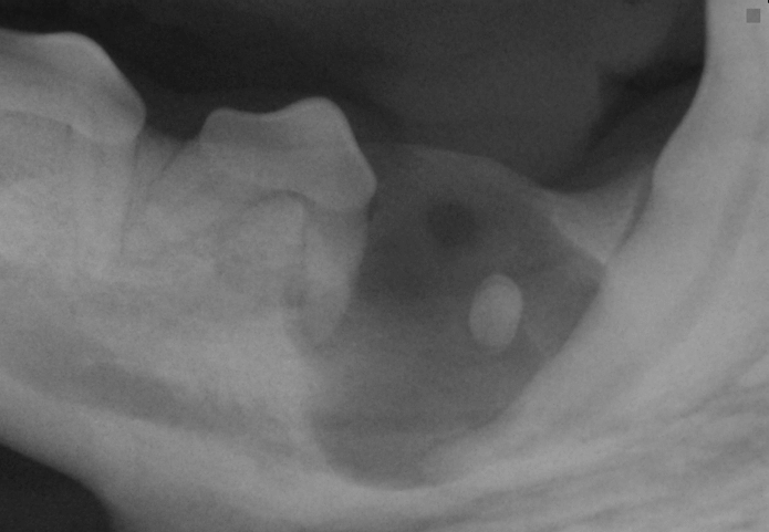
Missing teeth that are truly missing are fine, but teeth that appear missing because they are not seen but exist below the gumline are problems. When this tooth does not erupt, the cells continue to try to produce parts of the tooth creating fluid. This happens gradually and the fluid pressure within the jaw causes resorption of the bone around the tooth. The area continues to fill with fluid as the bone gradually resorbs resulting in little to no support 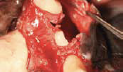until the jaw breaks. All sites of missing teeth need to be verified by intraoral x-rays to see if truly missing. If the cyst (described above), called a dentigerous cyst, has formed, it needs to be removed along with its lining so the cyst does not recur. Many cases have significant bone loss requiring grafts and/or membranes to facilitate appropriate healing.
The above image explains dentigerous cysts well. It is important to remember unerupted teeth and retained tooth roots both are likely to cause problems. If you notice a tooth that does not have a matching tooth on the other side, make sure to ask your veterinarian to check it with intraoral x-rays. Should you own a breed with a short nose aka brachycephalic, missing teeth on both sides is more likely a problem thus may be easily overlooked. Please ensure your dog has full mouth intraoral x-rays or [3D imaging](https://www.fampetvet.com/services/3-d-imaging) as it is shown intraoral x-rays reveal unexpected or additional new findings in 72% of dogs.
May be seen visually:
While an x-ray may look like this:
- #### Enamel defects 🐕🦺
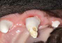Trauma, drugs, or disease during tooth development can result in abnormal enamel that does not provide good protection to the underlying dentinal tubules eventually resulting in internal disease. Special multi-step products are used to prepare and fill in the defect limiting the damage and improving the cosmetic appearance.
The above image explains enamel defects well.
Some cases of enamel hypomineralization appear as discoloration. While this is not only cosmetic, it is important to help seal any access to the central aspect of the teeth.
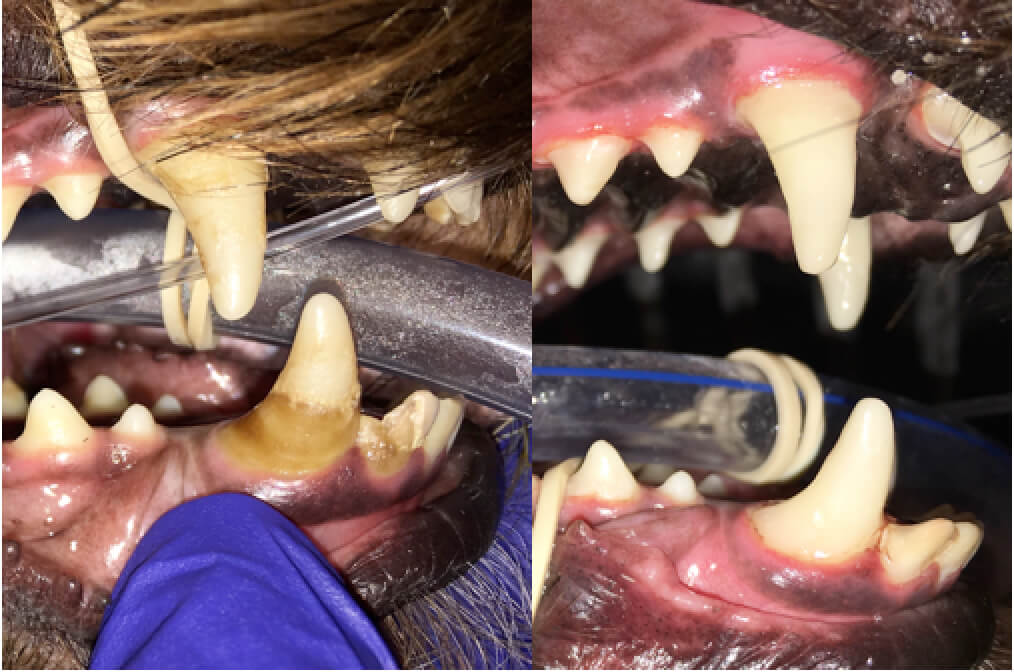
Other cases have focal defects.
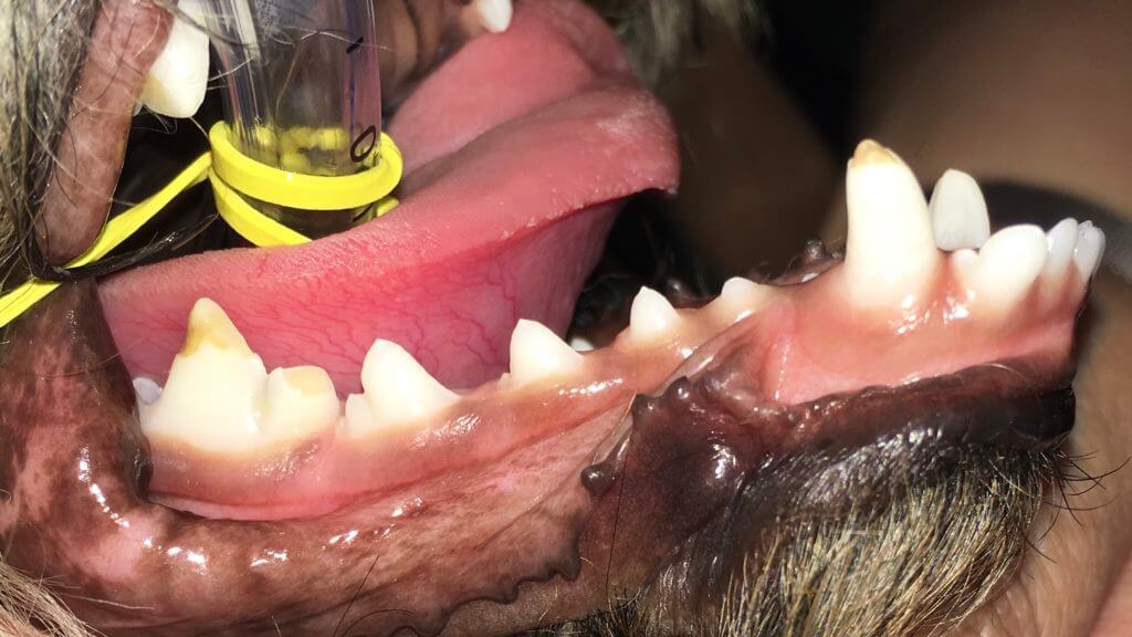
Once the defects have been sealed, the restorations will wear away as they are not as strong as natural enamel and there are inherent defects in the patient’s underlying tooth quality. In most cases, restorations can and should be reapplied.
Just as noted in other reasons for restorations such as caries and uncomplicated crown fractures, x-rays are needed before a restoration is applied and 6 months later. These patients, as well as every pet, should have intraoral x-rays with preventative cleaning procedures at least every 12 months.
- #### Tissue regeneration 🐕🦺 🐈
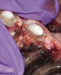Some cases of periodontal disease results in bone loss that still has surrounding and supporting bone. These are areas for which bone grafts and/or special membranes should be used to perform guided tissue regeneration to save a tooth.

As the gum and bone surrounding the tooth margin (periodontium) have worsening poor health, [extractions](https://www.fampetvet.com/services/extractions/) are often needed. These periodontal tissues usually have generalized disease and accompanying bone loss resulting in loss of tooth support. Other times the tooth has focal disease for which therapy can be successful in saving the tooth.
Vertical bone loss is a case in which deep pockets exist with supporting bone. A standard dental cleaning procedure cannot adequately clean deeper than 5 mm. The other concern with pockets greater than 3mm is that there is gingival down growth commonly reestablishing active disease as soon as 2-14 days after a cleaning procedure. Home care (daily or every other day) and anesthetic dental procedures (as often as every 3 months) can delay this process if it can get into the pocket but the pocket depth often prevents it. The solution becomes to surgically reduce the soft tissue pocket or, as pictured above, utilize a bone graft and biocompatible membrane to exclude the soft tissue from a bony pocket. The membrane is the most important feature to prevent tissue downgrowth perpetuating a pocket.
Here’s a hidden pocket:
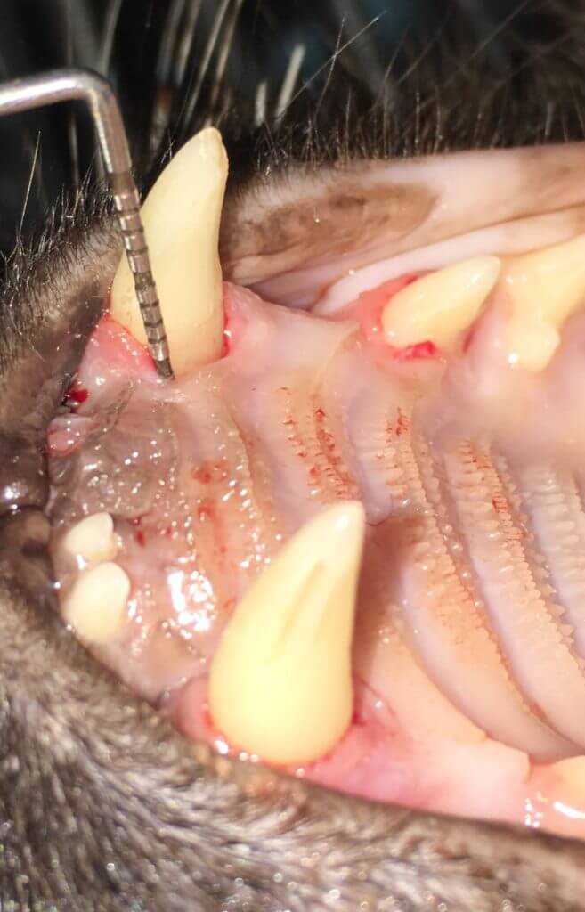
Here’s the diseased tissue in the pocket removed:
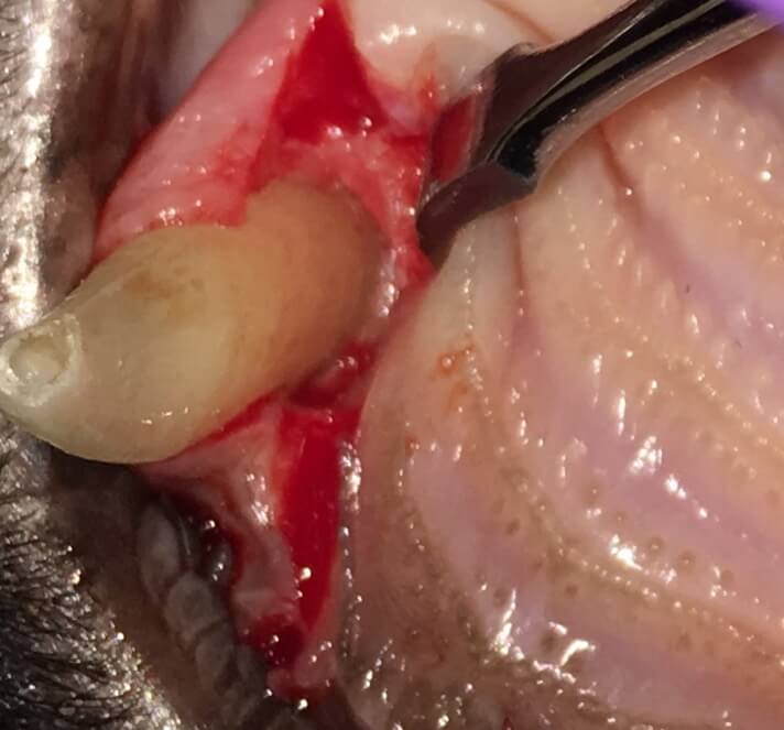
A graft and membrane are needed/were used.
- #### Oral nasal fistulas 🐕🦺 🐈
Some types of bone loss around teeth results in the need for flaps to repair the area around the tooth or close the defect in the area of the missing tooth. It is important to treat these correctly or the disease will return in as little as two weeks. Some of the major areas we treat are under-erupted lower canines, triangular gingival defects, and oral nasal/antral fistulas (ONF/OAF). ONFs and OAFs are openings between the mouth and the nasal sinuses.
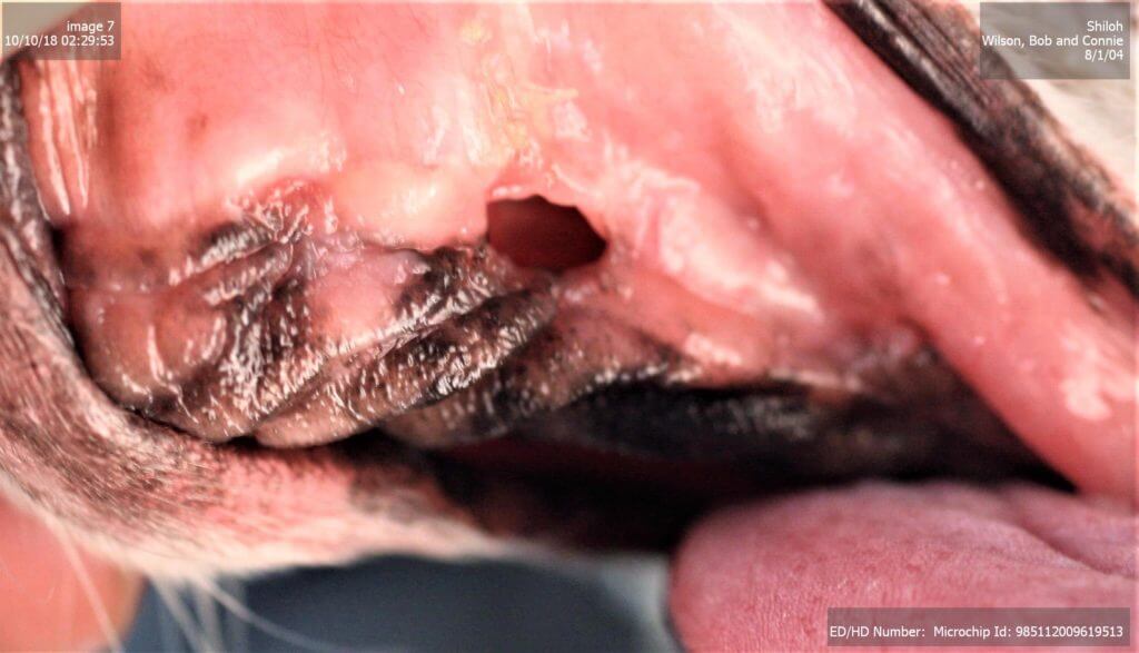
Sometimes these openings are large or easily seen as in the above images. Sneezing and occasional discharge are the common signs. Other times signs are subtler but systemic (body wide) effects are occurring without awareness.
Here is an inapparent oral nasal fistula:
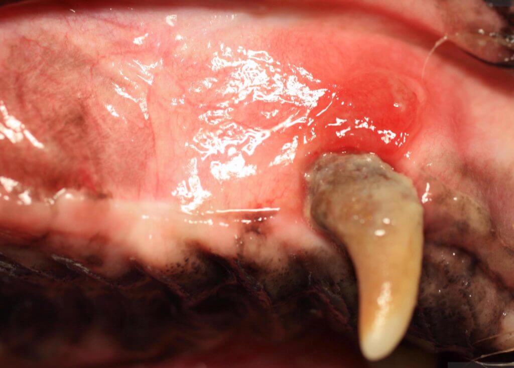
They are not always this red.
This one is easier to guess an oral nasal fistula is present:
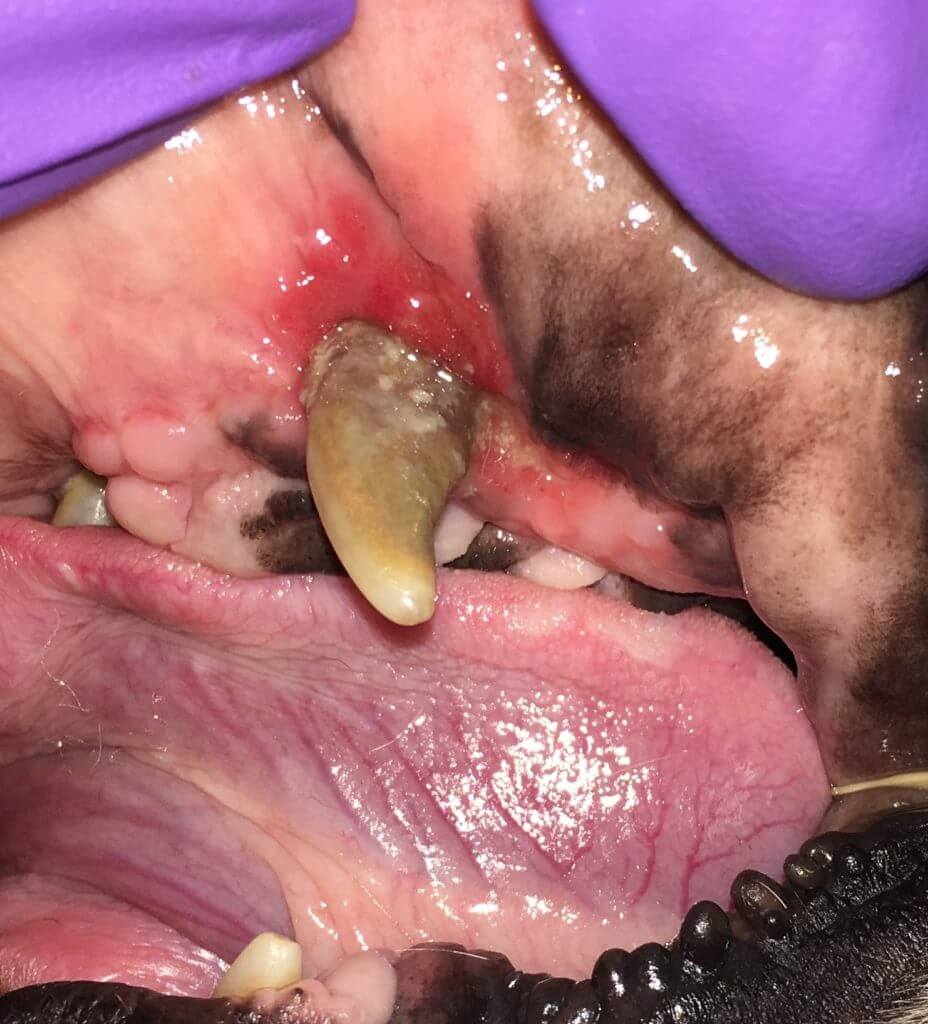
Tooth removed:
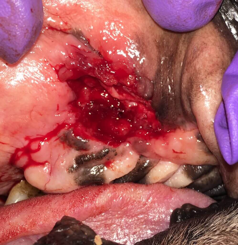
Unfortunately one can see the tissue quality is poor and doesn’t close easily. This is why the opening often happens. Special tissue flaps are created to close the defect to stop the communication between the nose and the mouth thus stopping subsequent infections. During veterinary dentistry residency training, we learn the periodontal classification as PE3 – a major flap procedure. Ie simple closure rarely is the solution due to local infection, tissue quality, and tissue quantity.
This video isn’t the best illustration but there is bone infection (osteomyelitis) and special techniques are need to close the defect:
Special flaps can be done for periodontal splinting, increased pocket depths, triangular gum recession – and much more. As long as there is sufficient healthy bone, we can save teeth with special flaps. Sometimes we can even add bone height with bone augmentation (guided tissue regeneration section) or Type II crown lengthening procedures. When teeth can not be saved, we have special flaps and techniques to return the mouth to a healthy state and avoid bone infection.
- #### Gingival hyperplasia 🐕🦺
Areas of enlarged gum tissue can be present, related to genetics or drug reactions, while other times it is related oral tumors. In the case of non-tumor causes, the problem is likely called gingival hyperplasia. This creates pockets between the tissue and the tooth trapping food, hair, and debris. The gums need to be resected to normal levels, removing the pocket. In many cases they will re-grow, so follow-up is necessary to keep the pet comfortable and prevent tooth loss secondary to periodontal pockets.
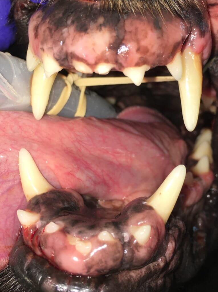
While the above images look drastic, this particular patient was much-improved 1 year later:
With the mild regrowth, retreatment is still needed, but there has not been destruction to the point of tooth loss. Left untreated the excess tissue traps food and hair between the tissue and the teeth resulting in loss of supporting structures and eventually teeth.
The above images and statements have been referring to gingival hyperplasia. Technically it cannot be said what this tissue is without biopsy results. We can say for certain is that there is gingival enlargement (GE). Whether the gingival enlargement is due to hyperplasia or hypertrophy, or whether the enlargement involves extensive thickening of the tissue, the goal is the same: return the structure and function of the area to a healthy sulcus (the space between the gum and the tooth).
Here are some additional examples of gingival enlargement before and after treatment.
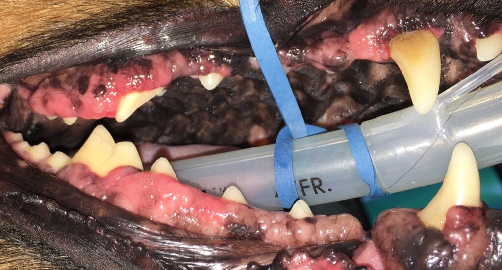
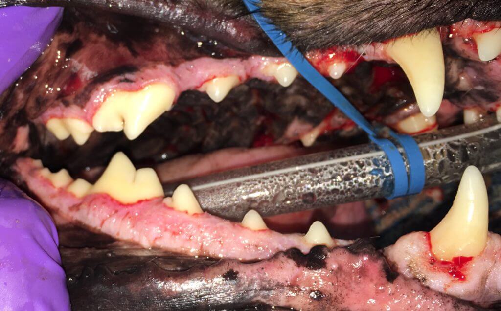
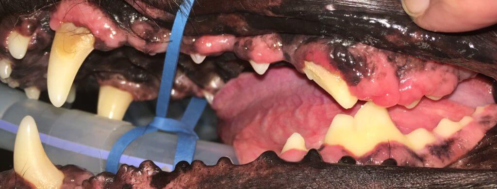
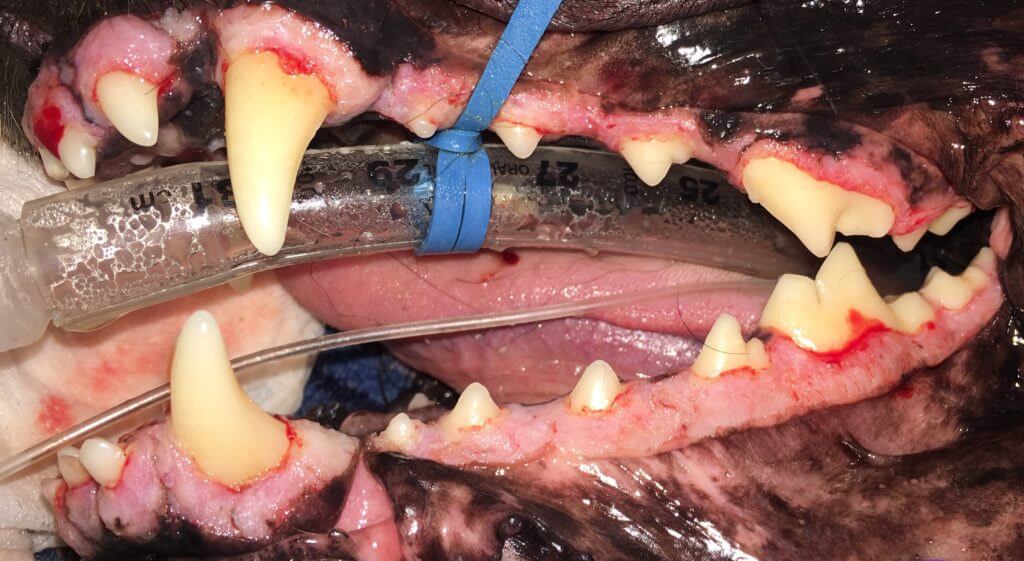
Though sometimes quite focal, often these lesions are generalized, particularly in breeds that show a familial tendency, such as Boxers, Bulldogs, and Collies.
Gingival enlargement can also be caused by hormonal changes or different drug administration (e.g., cyclosporine, amlodipine) in some patients.
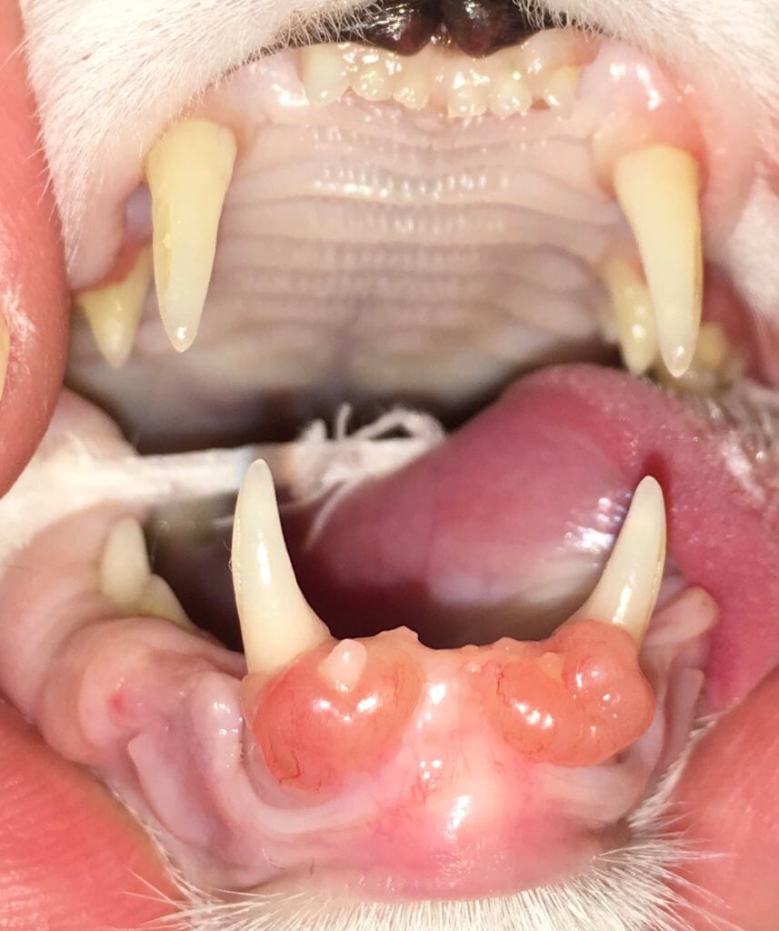
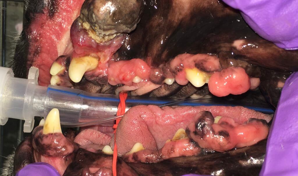
Some gingival enlargements may actually be growths or tumors and seem like other non-concerning changes seen in other patient. This is why all types of GE should be biopsied. Biopsy samples should be sent to an ORAL pathologist. Dr. Cindy Bell (KSU) currently is the leader oral pathology. Speaking from experience, it appears her reads tend to be more definitive than others. She is down to earth and will actually talk to veterinarians about treatment or help provide connections to speak to a veterinary dentist about the case.
Check out the [oral tumor](https://www.fampetvet.com/services/oral-tumors/) section to learn more.
- #### Oral tumors 🐕🦺 🐈
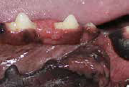Many types of oral tumors exist. Some are benign but locally painful and aggressive. Others are malignant but can still be treated with a high percentage of success. All gum enlargements should be biopsied or resected and submitted to a special oral pathology lab as many similar-looking masses can be many different results necessitating different additional therapies. Studies show 32-100% (depending on tumor type) remission with 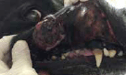wide-margined surgical excisions. Most pets respond well to these major surgeries and live a quality life enjoying further companionship.
While only 6% of malignant tumors of the body occur in the mouth, additional growths classified as benign occur. The trouble with the benign classification is that although they may not spread to other parts of the body, they are often locally invasive, painful, and destructive. The other trouble with both benign, benign-locally invasive, and malignant tumors is that many types of them look quite similar. As stated in the prior section, all types of gingival enlargements should be biopsied to help ensure the best treatment. Biopsy samples should be sent to an ORAL pathologist. Dr. Cindy Bell (KSU) currently is the leader oral pathology. Speaking from experience, it appears her reads tend to be more definitive than others. She is down to earth and will actually talk to veterinarians about treatment or help provide connections to speak to a veterinary dentist about the case.
Classifications of the following image growths may be plasma cell tumor, peripheral odontogenic fibroma, focal fibrous hyperplasia, papillary squamous cell carcinoma, lymphoma, pyogenic granuloma, canine acanthomatous ameloblastoma. I’m sure there are a few I’ve missed as well. Take note – the word epulis was not used to describe any of the growths. Epulis is a poor descriptive term. Most previously classified epulides are either peripheral odontogenic fibromas or ossifying fibromas. Also note, that all of the below images look similar and most of them are severe, locally invasive, or symptoms of additional disease.
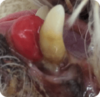
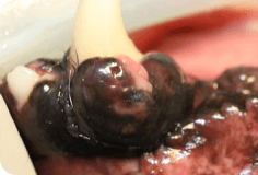
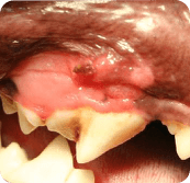
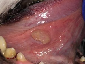
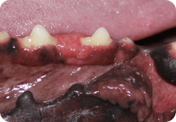
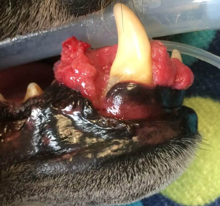
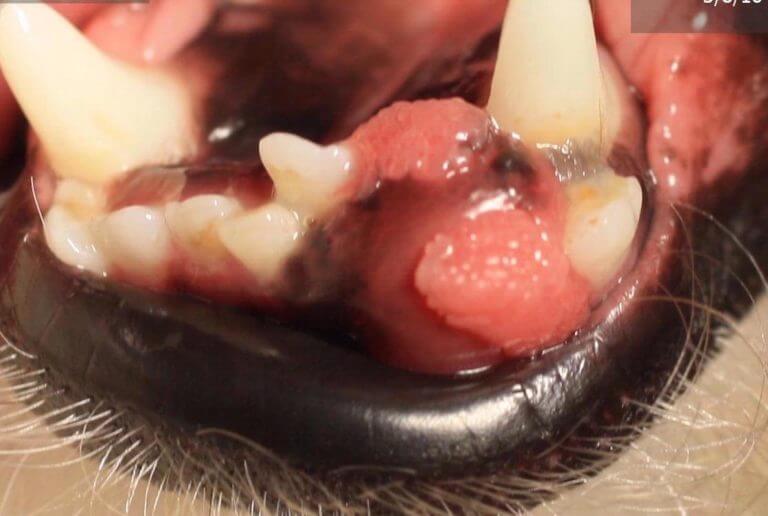
Many of these tumors require wide margined surgical excision; meaning large parts of the jaw(s) are removed. When taking off parts of the upper jaw, large holes can be made but the lip can cover and make the outcome be quite cosmetic for many cases.
A mandibulectomy finish is shown below.
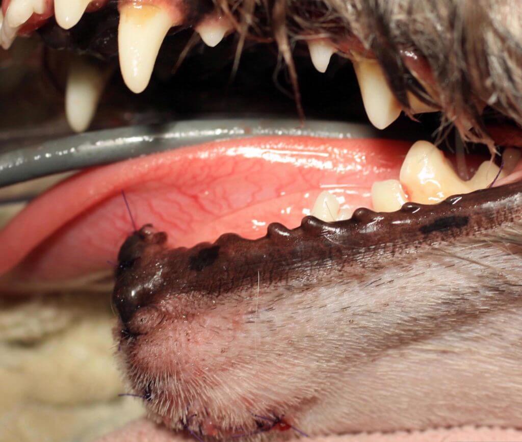
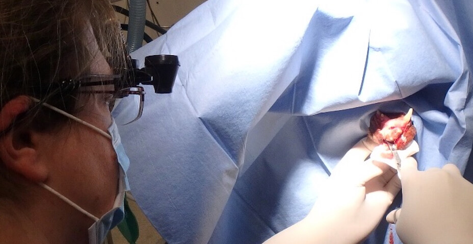
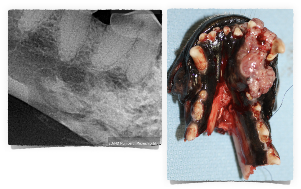
Studies show client satisfaction following mandibulectomies and maxillectomies in dogs is 85%, and was proportional to the postoperative survival time. It has been thought that cats do not do well after these types of surgeries. Surprising to many, a similar percentage of clients with cats after a mandibulectomy procedure said they would choose the same course of action given the circumstance despite the fact that 75% of the cats had mild to moderate adverse effects (tongue location, drooling, difficulty grooming, jaw alignment issues) for the rest of their lives.
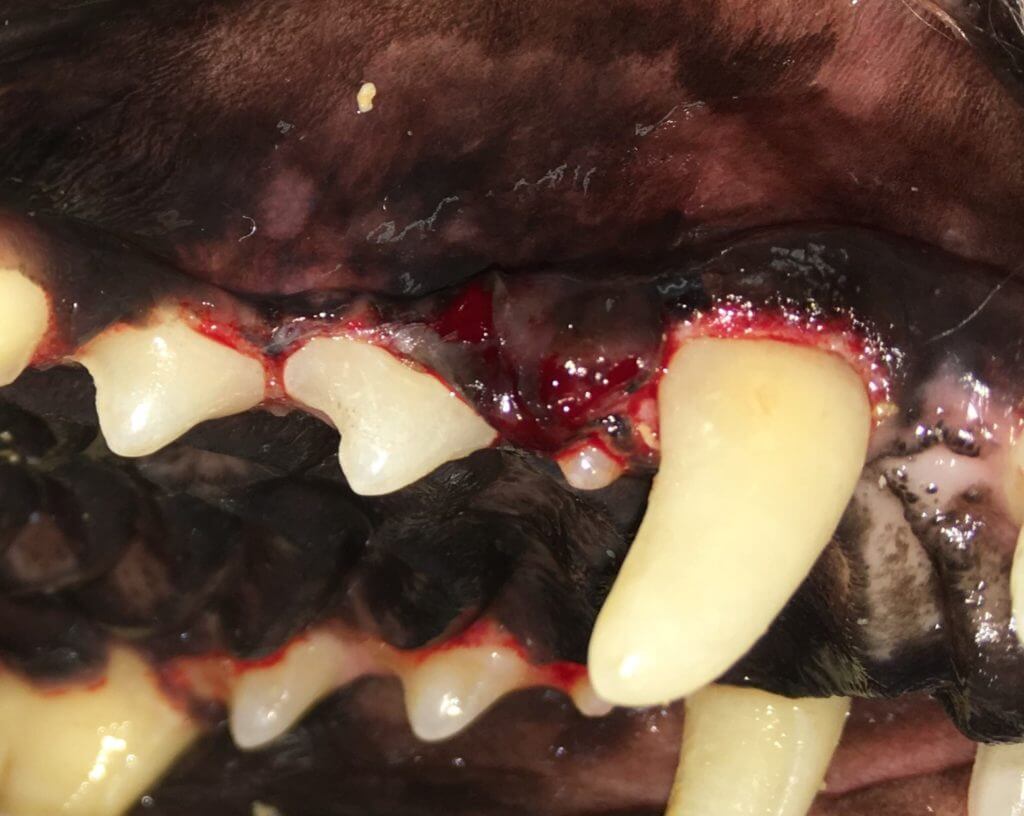
This mass (behind the canine and is subtle) was found very early and had a great outcome. Other larger ones of this type usually do better with adjunctive radiation therapy. Some masses even if found later in the course of disease have great surgical outcomes. For oral tumors it has been shown that all do much better with wide margined surgical excision with or without chemo therapy/radiation as compared to chemo therapy/radiation without surgical excision.
Determining surgical margins is important. 3D imaging with contrast can help make this determination in some cases. Read more about our high definition volumetric imaging capability with 0.09mm detail here. Information on 3D imaging of exotics and imaging of nasal issues can be found here.
##### Quick Links
Quick Links
- [Refer a Patient](https://tooth.vet/patient-referral)
- [Schedule a Consultation - For Pet Owners](https://tooth.vet/pet-owners)
- [Dental Education](https://tooth.vet/dental-education)
[/Animal-Dentistrylogo-transparent.png)](https://tooth.vet/about-us?hsLang=en)
© 2025 Animal Dentistry Referral Services
[Facebook](https://www.facebook.com/FamilyPetNorwalk)[YouTube](https://www.youtube.com/@toothvetia)
Return to top